INTRODUCTION
Lumbar spinal stenosis (LSS) is a well-known cause of low back pain and neurogenic claudication which cause disability, especially in aged people. Nonsurgical conservative treatments including activity modification (core muscle strengthening exercise, posture change, and restriction of activity), medication, epidural steroid injections are helpful for patients with mild to moderate symptoms. If conservative treatments fail, surgical intervention will be considered as the next treatment. Surgical decompression for LSS is indicated in a patient with intractable pain, neurogenic claudication, and motor weakness, accompanied by compromised spinal canal.
Conventional laminectomy is the most common surgical approach for decompression of spinal stenosis, but it may cause fibro-muscular and bony structure damage leading to significant instability, which results in poor long-term prognosis. And extensive resection of the structures can be associated with extensive perioperative blood loss, prolongation of postoperative pain, and the delay of hospital stay19). There are many technical modifications for decompression to relieve the problem associated with the conventional laminectomy3,5,8,21,26).
Central decompressive laminoplasty (CDL) was introduced as one of the modifications of the decompressive surgery. In the previous study, the author reported the surgical techniques and early surgical outcome15). The aim of this study was to present the long-term clinical and radiological outcomes of CDL for the treatment of LSS.
MATERIALS AND METHODS
Between December 2010 and March 2014, 100 patients with LSS underwent CDL by a single surgeon (YJK). The patients underwent consistent baseline clinical and radiological investigation, and adequate follow-up over 12 months was possible in 68 patients. Most patients have low back/buttock and leg pain, and/or claudication. All patients underwent adequate conservative treatment including pain medicine, physical therapy, and nerve block for over 6 weeks before the operation. LSS was identified on magnetic resonance imaging and/or computed tomography (CT) with 3-dimensional reconstruction. Preoperative dynamic X-rays were taken of all patients to diagnose significant spinal instability. LSS accompanied with herniated disc, or grade 1 degenerative spondylolisthesis was also included. Patients who had a high grade (over grade 2) spondylolisthesis and stenosis accompanied with the traumatic or neoplastic disease were excluded from this analysis.
The Institutional Review Board of Kangbuk Samsung Hospital approved the study protocol (KBSMC 201707026). Informed consent was waived by the board.
1. Outcome Assessment
The pain was measured separately for back/buttock and leg according to a patient-reported outcome with a 10-point visual analog scale (VAS). Functional disability was checked using the Oswestry Disability Index (ODI), which has been culturally validated for Korean-language speakers10).
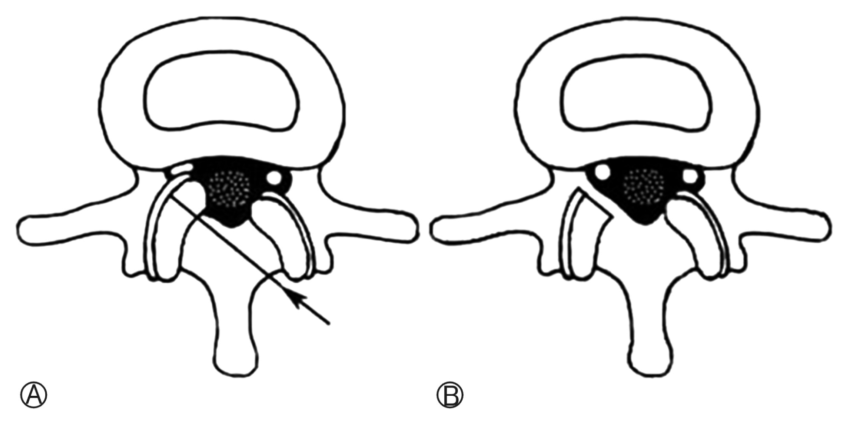
Radiological parameters are assessed by lateral X-ray on neutral position and dynamic posture with flexion and extension. The parameters were evaluated to determine the progression of spinal instability. Progression of spondylolisthesis was assessed on neutral and dynamic position. It was calculated by neutral slip percentage, the slippage divided by the length of the upper line at the lower vertebra, and dynamic slip percentage, the difference in the slip percentage between the flexion and extension posture. The dynamic intervertebral angle was calculated by the difference in intervertebral angles during flexion and extension (Fig. 1). Radiographs were analyzed using the PiView STAR program(INFINITT Healthcare, Seoul, Korea).
Outcomes after CDL were analyzed using changes of clinical and radiological parameters from the preoperative baseline. Postoperatively, the parameters were surveyed at 3 months, 6 months, 1 year, 2 years, 3 years, and thereafter. For the patients following more than 3 years, the last follow-up data were selected for analysis. An independent researcher conducted the survey at the time of follow-up. Data was collected prospective manner and analyzed retrospectively via electronic medical record and image software review.
2. Statistical Analysis
The mixed-effect model was used to test for differences from baseline in VAS scores of back/buttock, VAS scores of leg, and ODI measured repeatedly within a patient. Random effects were each parameter for patients, and error. Fixed effects were times at baseline and postoperative periods. Multiple comparisons between baseline and each follow-up time value were adjusted by Bonferroni method. Data were analyzed using the IBM SPSS Statistics ver. 24.0 (IBM Co., Armonk, NY, USA). A p-value of <0.05 was considered statistically significant.
3. Surgical Technique
Detailed surgical technique and related images were described in the previous study15). Brief technique is as follows. Under the general anesthesia, the patient was positioned on the Wilson frame, and surgical level checked using a C-arm fluoroscopy. After midline 3-cm-sized skin incision is performed over the spinous process and down to supraspinous and interspinous ligament. The sharp incision onto the ligaments enabled the functional soft tissue closure after primary operation. During bilateral dissection of the muscles exposing laminae of vertebrae, special care was done not to violate the facet capsule. After retraction of dissected muscle bilaterally, a small portion of the inferior spinous process of the cranial vertebra is removed, and the superior portion of the lamina in caudal vertebra is removed with a bone rongeur. The spinolaminar junction of the vertebrae is drilled with a cutting burr. The prone positioning with elevated curvature of the frame and release of ligamentum flavum from the lamina opens interlamina waking space. The ligament was removed with a rongeur. The capsular portion of the ligament flavum was not disturbed to maintain facet joint stability. Using a high-speed drill and spinal punches, undercutting of caudal lamina was performed.
Decompression of nerve roots was performed from the contralateral side using an operating microscope. Tilting down the operative table to opposite side provide the surgical corridor to remove upper proximal exiting root. This contralateral approach made possible the undercutting of the facet joint while decompressing distal traversing root. The facet was removed in a tapered fashion, which allowed complete decompression of the root and sparing of the facet capsule (Fig. 2). Complete decompression of the root was performed along the pedicle to the lowermost part of the recess, at the turning point of the root into the neural foramen. Discectomy was performed when soft herniated disc existed. The divided ligaments and skin were closed after an insertion of a drain.
RESULTS
Sixty-eight patients had adequate long-term clinical and radiological follow-up. Mean follow-up time was 37.7 months (range, 12–85 months). Average age at the time of surgery was 61.6 years (range, 31–81 years). Male to female ratio was 0.79 (male, 30; female, 38). CDL was done for 86 stenotic levels and L4/5 is the most frequent surgical level (51 levels, 59.3%). Discectomy was done for 23 patients (33.8%) and 25 levels (29.1%). Fifty of 68 patients (73.5%) were performed one-level surgery. Nine patients (13.2%) underwent previous lumbar spine surgery from other surgeons (Table 1).
No serious complications were noted during perioperative and early postoperative period. There were 3 additional surgeries after initial CDL during the follow-up. A 64-year-old woman who underwent CDL on L1/2 and L2/3, one and half years later, required discectomy on the right side of L1/2 after lifting trauma. The other 2 patients had received upper and lower adjacent root decompression for far-lateral foraminal stenosis after 3 years and 1 year/3 months after the initial surgery, respectively.
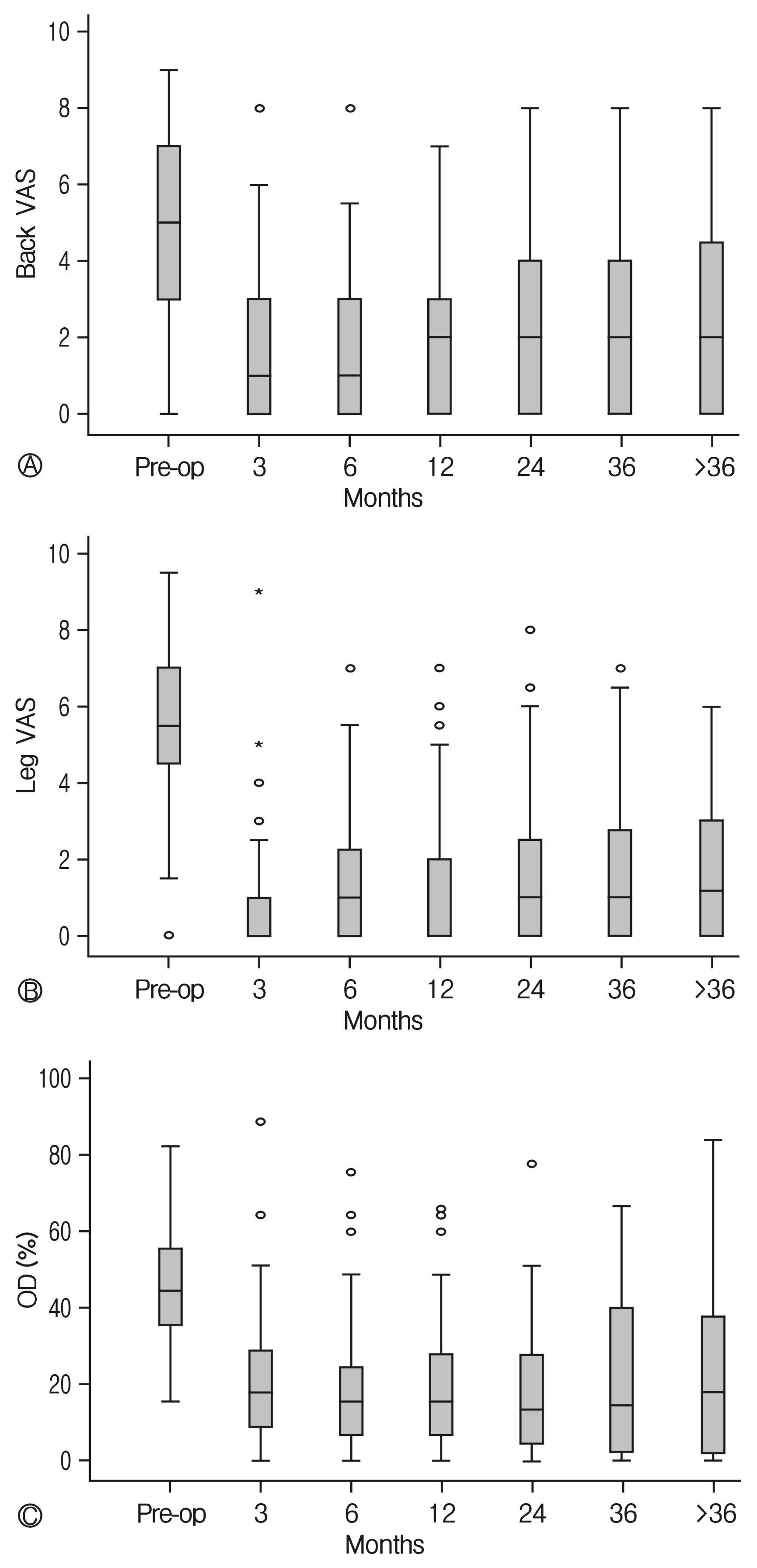
Mean preoperative VAS scores of back/buttock (backVAS), VAS scores of leg (legVAS), and ODI were 4.3 (median, 5.0; inter-quartile range [IQR], 2.8–7.0), 5.6 (median, 5.5; IQR, 4.5–7.1), and 45.0 (median, 44.4; IQR, 34.5–55.6), respectively. The 3-month follow-up data (backVAS; 1.7 [median, 1.0; IQR, 0–3.0], legVAS; 0.8 [median, 0; IQR, 0–1.1], ODI; 21.3 [median, 17.8; IQR, 8.9–30.0]) showed significant improvements vs. the baseline values (p<0.001, respectively). The mixed-effect model can analyze long-term trend of outcome after the surgery. It showed that back and leg VAS scores improved significantly in the early postoperative period (3 months) and maintained at plateau with time. ODI was lowest at 6 months postoperatively and tended to maintain plateau over the follow-up period (p<0.001, respectively) (Fig. 3).
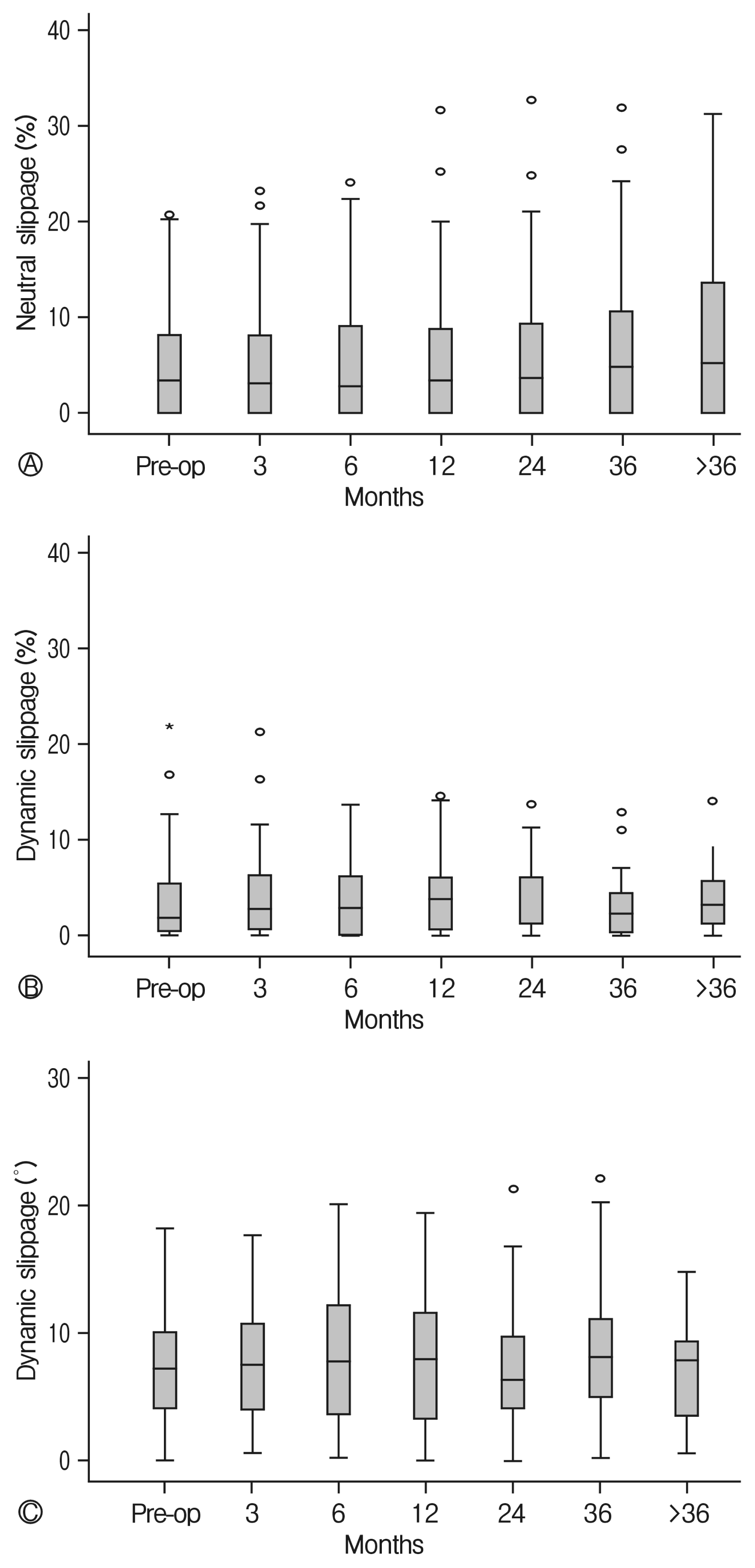
Baseline mean neutral slippage percentage was 4.9 (median, 3.8; IQR, 0–8.5), and the baseline preoperative dynamic slippage percentage (the difference in the slip percentage during flexion and extension posture) was 3.5 (median, 1.8; IQR, 0.5–5.4). Another tool for assessing dynamic instability, dynamic intervertebral angle, was checked mean 7.6° (median, 7.3°; IQR, 4.1–10.1) at baseline. During the post-operative follow-up, the operative levels were well maintained without significant progression of spondylolisthesis in neutral position (p=0.186). Dynamic slippage and dynamic intervertebral angle were not increased over the follow-up time (p=0.236 and p=0.123, respectively), which means good maintenance of spinal stability (Fig. 4).
DISCUSSION
Decompressive laminectomy has been known as a standard technique for treatment of degenerative LSS. Generally, the long-term prognosis of the technique has been reported poor as time passed by, despite wide variational and little data from long-term follow-up12,13,24). Some studies reported good long-term results using minimal resection of the facet joint during sufficient neural decompression5,22), suggesting preservation of biomechanical stability, especially in the facet joint area, is required for long-term success of this technique.
Conventional total laminectomy could provide wide neural canal decompression but leads to extensive resection of supraspinous and interspinous ligaments, spinous process, and lamina. And it needs wide removal of facet joints that provide spinal stability. Compromise of facet joints may transmit direct compressive forces to the disc and longitudinal ligament, and cause degenerative spinal instability7,18).
Several technical modifications of conventional laminectomy have been suggested to overcome this limitation. Bilateral laminotomy3,8) and unilateral laminotomy with bilateral decompression4,11,20,25) are well-known techniques. These methods enable surgeons to preserve midline structure while decompressing the-cal sac and nerve roots, and showed good surgical outcomes4,9). However, those have some technical shortcomings. First, preservation of midline supporting structure may interrupt the access to the ipsilateral lateral recess and foraminal area, especially if hypertrophied spinous existed. Next, large amount of facet resection is needed to access to the ipsilateral lateral recess and neural foramen, and violation of facet joint capsule occurs frequently. These may have an unfavorable effect on stability during the long-term follow-up. Recently, endoscopic surgery is another option of decompression for minimizing operation-related trauma. Although some authors published similar surgical outcome compared to previous microsurgical decompressive laminectomy14,17), the surgery is technically demanding in severe cases and has not enough data for safety and effectiveness16).
To overcome this limitation, we devised a specific modification of decompressive laminectomy, the CDL, which provide wide decompression while preserving facet stability by adequate bony resection limited in physiologic range15). Using the surgical corridor into the central interspinous area, the technique preserves near-total facet joint including the capsule overlying the joint. Excellent surgical view and unhindered route to the central canal, lateral recess, and upper/lower foraminal area provide thorough decompression of the neural elements. CDL provided an excellent early surgical outcome with significant pain and functional improvement.
In the present study, the author displayed long-term results of CDL for LSS using both clinical and radiological parameters. The mixed-effect model was used to adjust for missing data for the repeatedly checked data. The statistical model was useful in the retrospective analysis of this prospectively collected data with long-term follow-up. VAS scores for back/buttock and leg improved significantly from baseline in the immediate postoperative period (3 months) and maintained at plateau with time (p<0.001). ODI improved significantly immediate postoperative period and maintained during the follow-up (p<0.001) without worsening of the symptom. These results were comparable to other long-term clinical outcomes of standard modification of decompressive laminectomy, unilateral laminotomy with bilateral decompression4,9).
A systematic review for postoperative spondylolisthesis revealed that instability was seen in 12% of open laminectomy, and more frequently in pre-existing spondylolisthesis (12.6%)6). CDL provided stable radiological outcome showing no progression of instability during this long-term follow-up study. Three representative parameters for progression of spondylolisthesis; neutral slippage, dynamic slippage, and dynamic intervertebral angle23), were well maintained and not increased over the follow-up period. The facet joint and its capsule are key structures for spinal stability, which resist shear force (33%)1) and most of bending force (70%)2) in flexion position. As mentioned above, CDL preserves facet joint and capsule via inclined undercutting through the contralateral surgical trajectory. This could contribute to no progression of spondylolisthesis despite simple decompression without fusion.
This observational study of a long-term follow-up period, involved a relatively small number of patients. It is necessary to execute prospective controlled trials in a larger population with other laminectomy techniques (unilateral laminotomy with bilateral decompression, endoscopic laminotomy) or fusion surgery for confirmation of the current results.
CONCLUSION
CDL is a good surgical option for treating degenerative LSS. It can be easily applied, and allows excellent field visualization and decompression, sparing of ligament and bony structure to maintain the stability. It results in long-lasting pain relief and improved quality of life without progression of radiological instability.