Warning: mkdir(): Permission denied in /home/virtual/lib/view_data.php on line 81 Warning: fopen(/home/virtual/e-kjs/journal/upload/ip_log/ip_log_2024-04.txt): failed to open stream: No such file or directory in /home/virtual/lib/view_data.php on line 83 Warning: fwrite() expects parameter 1 to be resource, boolean given in /home/virtual/lib/view_data.php on line 84 Pneumocephalus and Pneumorrhachis due to a Subarachnoid Pleural Fistula That Developed after Thoracic Spine Surgery
Korean J Spine Search

CLOSE


Lee, Lee, Kim, Kim, Kim, and Kim: Pneumocephalus and Pneumorrhachis due to a Subarachnoid Pleural Fistula That Developed after Thoracic Spine Surgery

Abstract

Development of a communication between the spinal subarachnoid space and the pleural space after thoracic spine surgery is uncommon. Subarachnoid pleural fistula (SAPF), a distressing condition, involves cerebrospinal fluid leakage. Here we report an unusual case of SAPF, occurring after thoracic spine surgery, that was further complicated by pneumocephalus and pneumorrhachis postthoracentesis, which was performed for unilateral pleural effusion.

INTRODUCTION

Pneumorrhachis and pneumocephalus due to a fistula are rare. Another rare condition is a communication between the spinal subarachnoid space and pleural space after thoracic spine surgery. Pneumocephalus may manifest as severe headache4,5,7,9) and acute neurologic changes that mimic brain metastasis, seizure, or stroke. However, the physiology and management of pneumocephalus are not widely recognized. Here we report an unusual case of subarachnoid pleural fistula (SAPF), occurring after thoracic spine surgery, that was further complicated by pneumocephalus and pneumorrhachis postthoracentesis, which was performed for unilateral pleural effusion.

CASE REPORT

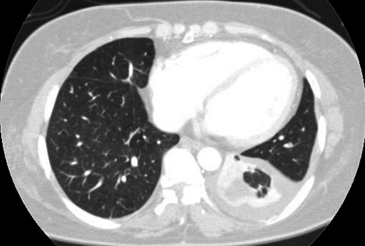
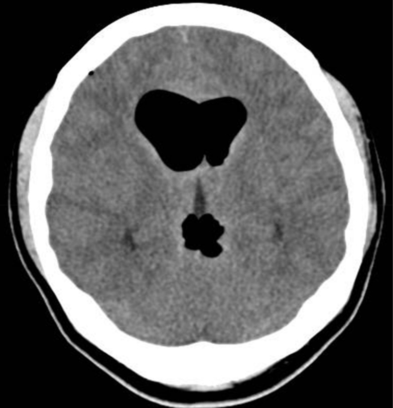
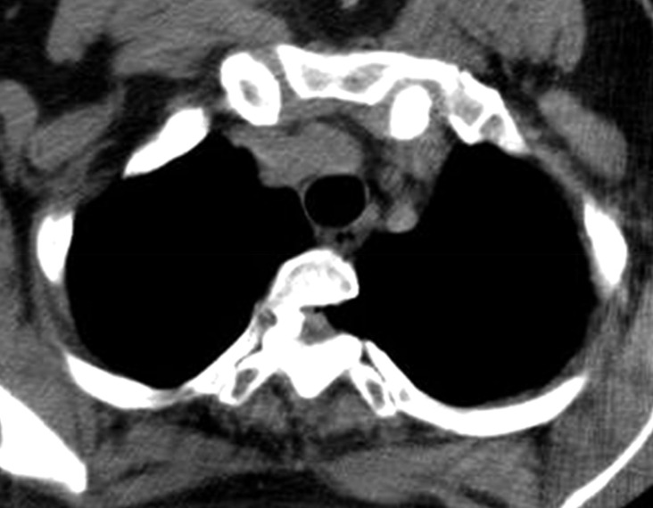
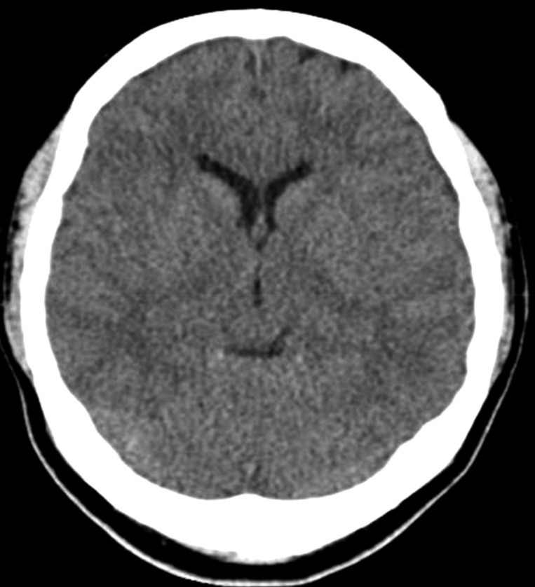
A 47-year-old woman presented with severe headache, nausea, and vomiting. A known case of T1-T8 posterior longitudinal ligament ossification, she had undergone decompressive laminectomy at T1-T2 and T8, 7 years ago and posterior decompression at T11-L2, 2 years ago at another hospital. At that time, dural tear was unknown. She was in her usual state of health until 10 days before admission to the respiratory department for violent coughing and sputum. A chest radiograph and computed tomography (CT) scan (Fig. 1) revealed left-sided pleural effusion. Thoracentesis was performed, following which she developed sudden headache, nausea, and vomiting. Brain CT scan revealed pneumocephalus, with air in the ventricle and the subarachnoid space (Fig. 2). A chest CT showed a communication between the pleural cavity and the T2 spinal canal with intraspinal air (Fig. 3). The precise location of the SAPF was identified on a CT myelogram with iohexol. The patient refused surgical repair of the SAPF; therefore, conservative management including bed rest with decubitus position was adopted as treatment. Her symptoms improved dramatically. A follow- up brain CT scan after 27 days showed complete resolution of the ventricular and subarachnoid air (Fig. 4). Consecutive chest radiographs showed left-sided pleural effusion, but she had no cough, difficulty in breathing, or sputum. She was discharged and sent home after 7 weeks since admission, and was followed-up in the outpatient department.

DISCUSSION

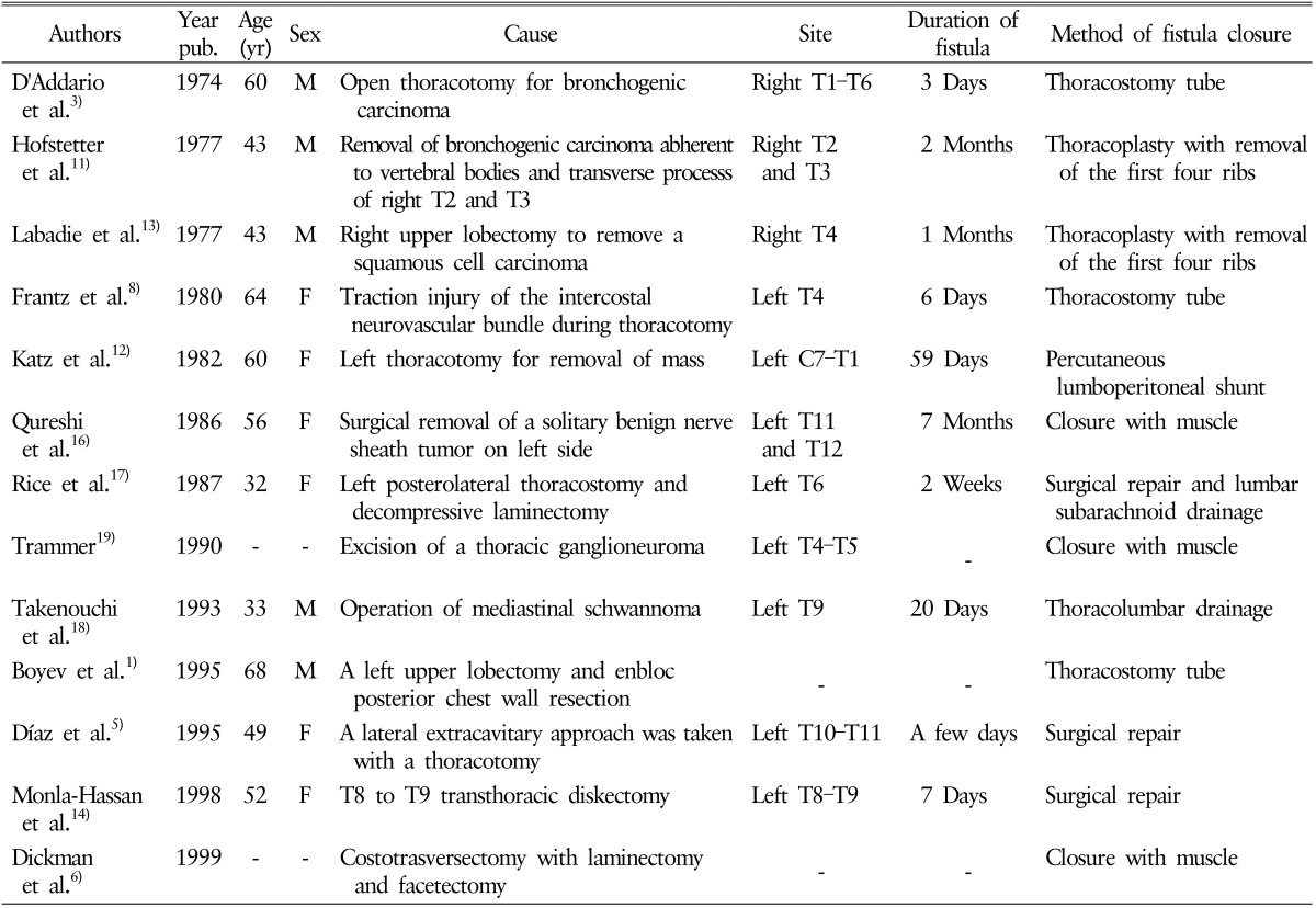
SAPFs are rare and difficult to diagnose. Only 13 cases of SAPF following surgery have been reported in the literature1,3,5,6,8,11,12,13,14,16,17,18,19) (Table 1). Postoperative pneumocephalus could result from combination of a dural tear and a pneumorrhachis. An SAPF may remain asymptomatic when small, or may cause pleural effusion and subsequent dyspnea and pleuritic chest pain, in addition to postural headaches4,5,7,9), nausea, and vomiting secondary to intracranial hypotension1). An SAPF once developed causes a pressure gradient, which allows cerebrospinal fluid (CSF) to flow from the positive-pressure spinal subarachnoid space to the negative-pressure pleural space. The low thoracic pressure created during inspiration causes CSF to flow into the chest cavity, and the high intrathoracic pressure during expiration forces air into the CSF space. This is further complicated by an upright head position that causes an increase in air in the subarachnoid space, resulting in pneumocephalus. The diagnosis of SAPF requires an appropriate clinical setting and relies mostly on the aforementioned symptoms and signs, although imaging modalities are of great value in confirming the presence of the fistula. The commonly used imaging modalities are radionuclide cisternography11), CT myelography10), and magnetic resonance imaging. Management of SAPF depends not only on the size of the fistula defect but also on the progression of symptoms, and choosing a treatment strategy is the most challenging part.
Several reports showing that conservative management is available in most of patients6,18).
In a review of 19 cases of SAPF secondary to trauma, 63% were treated surgically, 26% via chest tube drainage or lumbar drainage, and 11% conservatively15). Furthermore, 27% of the fistulae developing postthoracotomy closed spontaneously2). Conservative therapy, which includes bed rest, external lumbar drainage, and chest tube insertion, has been advocated as the initial treatment strategy. Surgical repair of the fistula should be considered after the failure of conservative treatment; however, the appropriate timing for chest tube drainage or surgical intervention remains controversial. Early detection and appropriate therapy will decrease morbidity associated with this rare complication.

CONCLUSION

Clinicians must be aware of the possibility of SAPF in patients with unilateral pleural effusion who have undergone thoracic spine surgery. Conservative management may be feasible in patients with SAPF, and long-term follow-up is imperative.

NOTES

CONFLICT OF INTEREST: No potential conflict of interest relevant to this article was reported.

References

1. Boyev P, Krasna MJ, White CS, McLaughlin JS. Subarachnoid-pleural fistula after resection of a pancoast tumor with hyponatremia. Ann Thorac Surg 1995 60:683-685. PMID: 7677501.
crossref pmid
2. Brown WM 3rd, Symbas PN. Pneumocephalus complicating routine thoracotomy: symptoms, diagnosis and management. Ann Thorac Surg 1995 59:234-236. PMID: 7818338.
crossref pmid
3. D'Addario R, Greenberg J, O'Neill TJ, Spagna P. Pneumocephalus: an unusual cause. J Neurol Neurosurg Psychiatry 1974 37:271-274. PMID: 4829533.
crossref pmid pmc
4. Da Silva VF, Shamji FM, Reid RH, del Carpio-O'Donovan R. Subarachnoid-pleural fistula complicating thoracotomy: case report and review of the literature. Neurosurgery 1987 20:802-805. PMID: 3037431.
crossref pmid
5. Díaz P, Maíllo A, Hernández J, Morales F, Jiménez MF, Moreta JA, et al. Subarachnoid-pleural fistula as a complication of the lateral-extracavitary approach to thoracic intraspinal neurinoma. Spine (Phila Pa 1976) 1995 20:1515-1518. PMID: 8623073.
crossref pmid
6. Dickman CA, Rosenthal D, Regan JJ. Reoperation for herniated thoracic discs. J Neurosurg 1999 91(2 Suppl):157-162.
crossref
7. Fernandez P, Guyot M, Mangione P, Valli N, Basse-Cathalinat B, Ducassou D. Subarachnoid-pleural fistula complicating thoracoscopy: value of In-111 DTPA myeloscintigraphy. Clin Nucl Med 1999 24:985-986. PMID: 10595487.
crossref pmid
8. Frantz PT, Battaglini JW. Subarachnoid-pleural fistula: unusual complication of thoracoscopy. J Thorac Cardiovasc Surg 1980 79:873-875. PMID: 6246314.
crossref pmid
9. Godley CD, McCabe CJ, Warren RL, Rosenberg WS. Traumatic subarachnoid-pleural fistula: case report. J Trauma 1995 38:808-811. PMID: 7760414.
crossref pmid
10. Hicken P, Martin J, Hakanson S. Computed tomography demonstration of subarachnoid-pleural fistula. Can Assoc Radiol J 1990 41:222-224. PMID: 2207780.
pmid
11. Hofstetter KR, Bjelland JC, Patton DD, Woolfenden JM, Henry RE. Detection of bronchopleural-subarachnoid fistula by radionuclide myelography: case report. J Nucl Med 1997 18:981-983.

12. Katz SS, Savitz MH, Osei C, Harris L. Successful treatment by lumboperitoneal shunting of a spinal subclavicular fistula following thoracotomy. Neurosurgery 1982 11:795-796. PMID: 7162573.
crossref pmid
13. Labadie EL, Hamilton RH, Lundell DC, Bjelland JC. Hypoliquorreic headache and pneumocephalus caused by thoraco-subarachnoid fistula. Neurology 1997 27:993-995.
crossref
14. Monla-Hassan J, Eichenhorn M, Spickler E, Talati S, Nockels R, Hyzy R. Duropleural fistula manifested as a large pleural transudate: an unusual complication of transthoracic diskectomy. Chest 1998 114:1786-1789. PMID: 9872224.
crossref pmid
15. Pollack II, Pang D, Hall WA. Subarachnoid-pleural and subarachnoid-mediastinal fistulae. Neurosurgery 1990 26:519-525. PMID: 2181337.
crossref pmid
16. Qureshi MM, Roble DC, Gindin RA, Scudamore HH. Subarachnoid-pleural fistula. Case report and review of the literature. J Thorac Cardiovasc Surg 1986 91:238-241. PMID: 3945091.
crossref pmid
17. Rice TW, Kirsh JC, Schacter IB, Goldberg M. Simultaneous occurrence of chylothorax and subarachnoid pleural fistula after thoracotomy. Can J Surg 1987 30:256-258. PMID: 3607639.
pmid
18. Takenouchi N, Miura T, Otake S, Muraki S, Kusaka T, Fujimori M, et al. Cerebrospinal fluid fistula following an operation of mediastinal schwannoma: a case report. Kyobu Geka 1993 46:1152-1155. PMID: 8258925.
pmid
19. Trammer A. Persistent subarachnoid-pleural cerebrospinal fluid fistula following excision of a thoracic ganglioneuroma: case report. Z Kinderchir 1990 45:109-110. PMID: 2360366.
pmid
Fig. 1

Chest computed tomography scan showing pleural effusion.

kjs-13-164-g001.jpg
Fig. 2

Brain computed tomography scan after thoracentesis showing pneumocephalus.

kjs-13-164-g002.jpg
Fig. 3

Chest computed tomography scan after thoracentesis showing air collection in the T2 spinal canal and a fistula.

kjs-13-164-g003.jpg
Fig. 4

A follow-up brain computed tomography scan, obtained after 27 days, showing the disappearance of the pneumocephalus.

kjs-13-164-g004.jpg
Table 1

Case of subarachnoid-pelural fistular following surgery

kjs-13-164-i001.jpg


Editorial Office
CHA University, CHA School of Medicine Bundang Medical Center
59 Yatap-ro, Bundang-gu, Seongnam-si, Gyeonggi-do 13496, Korea
Tel: +82-31-780-1924  Fax: +82-31-780-5269  E-mail: support@e-neurospine.org
The Korean Spinal Neurosurgery Society
#407, Dong-A Villate 2nd Town, 350 Seocho-daero, Seocho-gu, Seoul 06631, Korea
Tel: +82-2-585-5455  Fax: +82-2-2-523-6812  E-mail: ksns1987@gmail.com
Business License No.: 209-82-62443

Copyright © The Korean Spinal Neurosurgery Society.

Developed in M2PI

Close layer
prev next