Ganglioneuroma of the Sacrum
Article information
Abstract
Presacral ganglioneuromas are extremely rare benign tumors and fewer than 20 cases have been reported in the literature. Ganglioneuromas are difficult to be differentiated preoperatively from tumors such as schwannomas, meningiomas, and neurofibromas with imaging modalities. The retroperitoneal approach for resection of presacral ganglioneuroma was performed for gross total resection of the tumor. Recurrence and malignant transformation of these tumors is rare. Adjuvant chemotherapy or radiation therapy is not indicated because of their benign nature. We report a case of a 47-year-old woman with a presacral ganglioneuroma.
INTRODUCTION
Ganglioneuroma is an uncommon benign tumor of neural crest origin which is mainly localized in the posterior mediastinum, retroperitoneum, and adrenal gland1,6). Presacral ganglioneuromas are extremely rare and known as predominantly occuring in women15). Patients are usually young and show neurological signs and symptoms when the tumor developing a significant size4,7). As imaging modalities have become more widely performed, the number of ganglioneuromas found has increased. However, it is very difficult to diagnose ganglioneuroma early in the evaluation of the lesion using image modalities only14). We report our experience with a case of presacral ganglioneuroma.
CASE REPORT
A 47-year-old woman presented to hospital with a month’s duration of intermittent flank and iliac fossa pain, abdominal discomfort, and tingling sensation on left thigh. She had a history of transvaginal myomectomy with symptoms of mild, temporary diarrhea and abdominal discomfort 3 years prior to admission. There was no specific family history. There were no palpable masses observed on abdominopelvic examination. Computed tomography (CT) scans of the abdomen and pelvis revealed a finely demarcated, oval shaped mass arising separately from the ovary on the right side of her pelvis (Fig. 1). Pelvic magnetic resonance (MR) images showed an elliptical tumor on the left side of the pelvis which were well enhanced by contrast (dimensions: 3.4 cm×2.4 cm×4.5 cm). The tumor was adjacent to the left sacrum and displacing the left sacral nerve roots, without any evidence of bony invasion (Fig. 2).
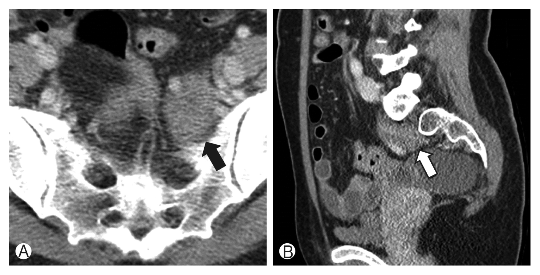
A pelvic computed tomography scan shows a well-demarcated and heterogeneous mass arising from the left S1 sacral foramen (arrow). Axial (A) and sagittal (B) view.
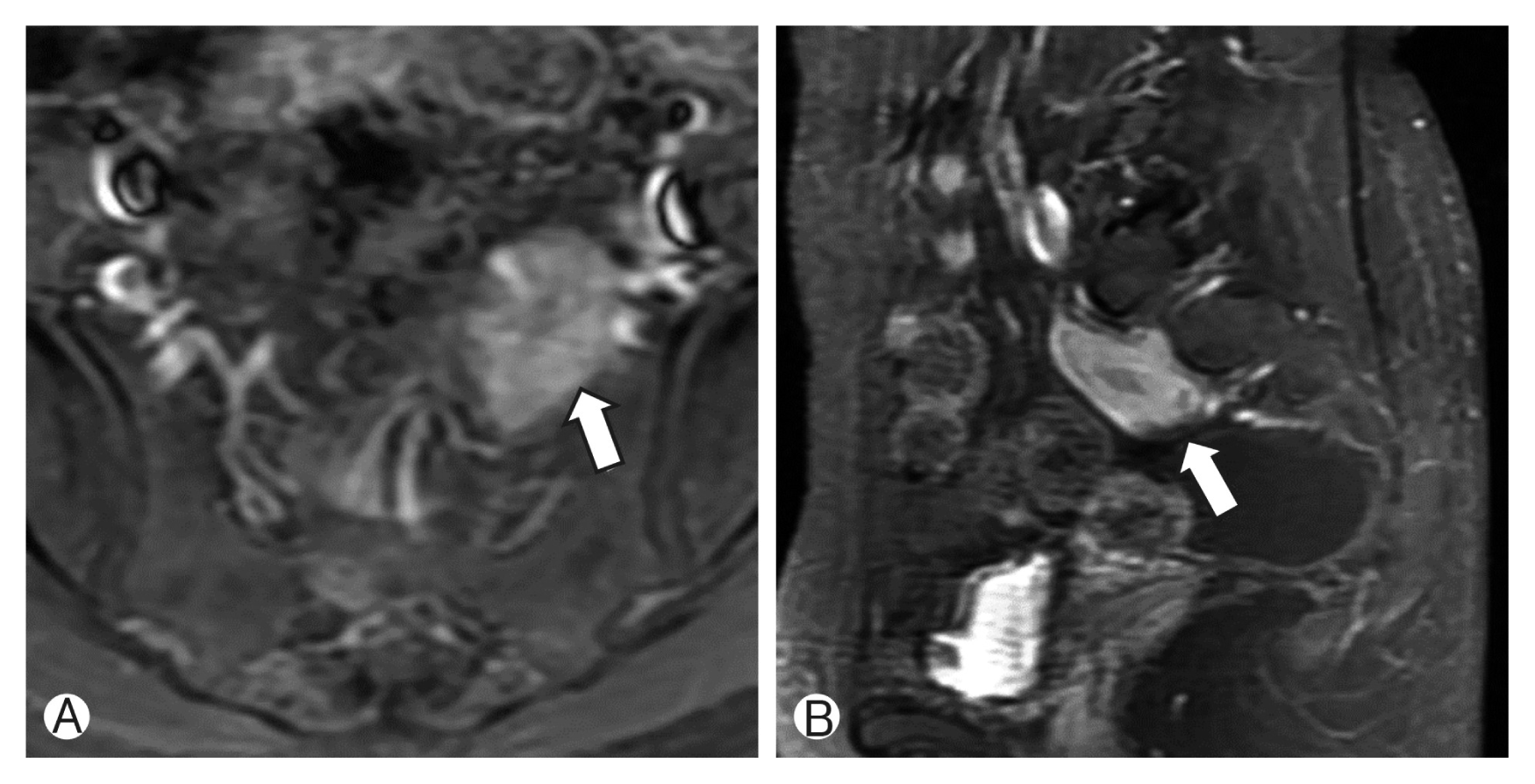
Magnetic resonance images show a presacral 3.4×2.4×4.5-cm mass (arrow). Contrast enhanced T1-weighted magnetic resonance image with intermediate high signal intensity. Axial (A) and sagittal (B) view.
We performed surgery via anterior retroperitoneal approach and meticulous adhesiolysis was necessary because of massive abdominal adhesion due to the previous gynecologic surgery. Especially, the left internal iliac vein and tumor capsule were adherent and were carefully dissected. We could discern the left S1 root using a nerve stimulator and the tumor was attached to the S1 nerve in the posterior side. Since the tumor was partly in the left S1 sacral foramen, the foramen was unroofed with drill and Kerisson punch to expose and remove the mass completely. The encapsulated retroperitoneal tumor located on the left side of the sigmoid colon was excised by piecemeal pattern and complete resection was performed (Fig. 3). Postoperative histopathologic study confirmed the lesion as a ganglioneuroma (Fig. 4). After the surgery, the patient recovered and discharged without any neurological deterioration.

Intraoperatively, a tumor is apparent after unroofing the left S1 sacral foramen.
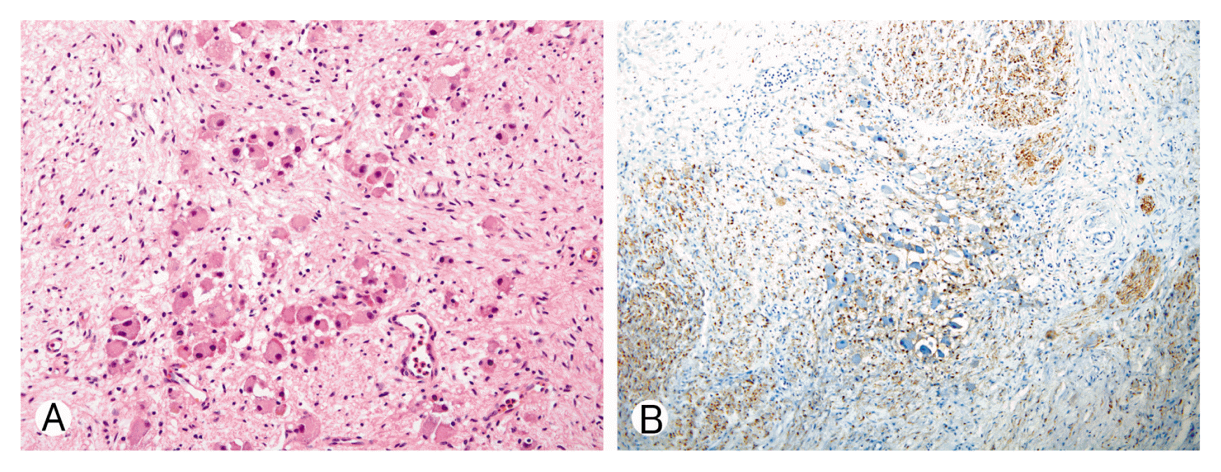
Histopathologic findings of ganglioneuroma. (A) Hematoxylin and Eosin staining with ×200 magnification. The specimen was composed of fibrous and edematous matrix with nests of ganglion cells and Schwann cells in the matrix. (B) S-100 and neurofilament staining at ×100 magnification.
DISCUSSION
Ganglioneuromas are well-differentiated, rare benign tumors of neural crest origin. Especially, presacral location of ganglioneuroma is extremely rare10,13,17,18). Ganglioneuromas often locate in the posterior mediastinum and retroperitoneum. The tumors usually show a slow-growing pattern with a predominance in women15). Most tumors are diagnosed at the progressed state of the tumor in patients between 10 and 30 years of age because symptoms only appear when the mass becomes large enough to exert a mass effect4,7). Ganglioneuromas can occur anywhere in the autonomic nervous system and can cause a mass effect on the spinal nerves and sympathetic nerves, leading to neurological abnormalities such as back pain, neurogenic bladder, urinary frequency, constipation, and bowel obstruction. Spontaneous or radiotherapy-induced malignant transformation of ganglioneuromas has been reported but this is extremely uncommon2).
According to our literature search, fewer than 20 cases of presacral ganglioneuroma have been reported worldwide. Ganglioneuromas arise from sympathoblasts derived from the embryonic neural crest. Histologically, ganglioneuromas consist of Schwann cells with mature ganglion and are considered to be part of the neuroblastoma group, along with neuroblastomas and ganglioneuroblastomas3,9,10,13,14,19). Preoperative evaluation could be performed by using fine-needle aspiration biopsy (FNAB), but the approach is often technically difficult because of the anatomical location. Also, using a single FNAB sample can lead to inaccurate diagnoses and cells should be sampled at multiple sites within the tumor8).
It is not easy to differentiate between ganglioneuroma and other tumors (such as schwannoma), only with clinical or imaging findings. There is no case report that patient was diagnosed with ganglioneuroma preoperatively with imaging modalities14). Magnetic resonance imaging (MRI) is a tool that not only helps to assess the nature of the lesion but also helps in planning of the surgery.
CT scans of ganglioneuromas may have dot-like calcifications in two-thirds of cases (this was not seen in the reported case), which can be a clue for differential diagnosis with neuroblastomas11). MRI helps in planning treatments by distinguishing lesions of spinal origin from lesions of pelvic origin14,16). On MR images, ganglioneuromas may show a high signal intensity during late enhancement and a heterogeneous mass on T2-weighted image. The structural and morphological characteristics of ganglioneuromas, such as the presence of mature sympathetic ganglion cells, can help distinguish them from other pelvic benign tumors such as schwannomas, meningiomas, or neurofibromas4,6,7,11). Appropriate therapy for ganglioeneuromas is complete surgical excision2,5). The complete resection of presacral ganglioneuroma provides for both effective diagnosis and treatment. Adjuvant chemotherapy or radiation therapy is not indicated due to the nature of this benign disease12).
In our case, an anterior retroperitoneal approach was used, which is a effective method for mass removal of presacral lesions because it provides a sufficient field of view to identify the origin of the lesion. This approach provided excellent access to the lesion and facilitated complete resection. Other groups have also reported that the posterior transsacral approach was proposed to remove a tumor completely involving the sacral nerve root. Modha et al.14) prefer the anterior approach, but a staged operation of laminectomy and foraminotomy can be added to manage any radicular symptoms after removal of the tumor.
Recurrence and malignant transformation of this tumor have been rarely reported2). In that case, the only treatment available is surgical resection. The patients’ prognosis is usually good and there is no consensus as to a follow-up plan.
CONCLUSION
Ganglioneuromas could be considered as a differential diagnosis in the treatment of presacral tumors. Anterior retroperitoneal approach is efficient method in complete removal of the tumor without neurological deficit.
Notes
CONFLICT OF INTEREST
No potential conflict of interest relevant to this article was reported.