Warning: mkdir(): Permission denied in /home/virtual/lib/view_data.php on line 81 Warning: fopen(/home/virtual/e-kjs/journal/upload/ip_log/ip_log_2024-04.txt): failed to open stream: No such file or directory in /home/virtual/lib/view_data.php on line 83 Warning: fwrite() expects parameter 1 to be resource, boolean given in /home/virtual/lib/view_data.php on line 84 Degenerative Cervical Myelopathy; A Review of the Latest Advances and Future Directions in Management
Neurospine Search

CLOSE


Neurospine > Volume 16(3); 2019 > Article
Wilson, Badhiwala, Moghaddamjou, Martin, and Fehlings: Degenerative Cervical Myelopathy; A Review of the Latest Advances and Future Directions in Management
See commentary "Degenerative Cervical Myelopathy" in Volume 16 on page 793.

Abstract

The assessment, diagnosis, operative and nonoperative management of degenerative cervical myelopathy (DCM) have evolved rapidly over the last 20 years. A clearer understanding of the pathobiology of DCM has led to attempts to develop objective measurements of the severity of myelopathy, including technology such as multiparametric magnetic resonance imaging, biomarkers, and ancillary clinical testing. New pharmacological treatments have the potential to alter the course of surgical outcomes, and greater innovation in surgical techniques have made surgery safer, more effective and less invasive. Future developments for the treatment of DCM will seek to improve the diagnostic accuracy of imaging, improve the objectivity of clinical assessment, and increase the use of surgical technology to ensure the best outcome is achieved for each individual patient.

INTRODUCTION TO DEGENERATIVE CERVICAL MYELOPATHY: A NEW OVERARCHING CONCEPT

Degenerative cervical myelopathy (DCM) is the most common etiology of spinal cord dysfunction among adults globally [1]. As an overarching clinicopathological entity, DCM encompasses a host of degenerative conditions of the cervical spinal column, including osteoarthritic degeneration (i.e., spondylosis) and ligamentous aberrations (i.e., ossification of the posterior longitudinal ligament [OPLL], ossification of the ligamentum flavum), that culminate in chronic compression of the cervical spinal cord, neural tissue destruction, and ultimately clinical loss of functional ability [2,3]. Even though cervical spondylotic myelopathy (CSM) and myelopathy secondary to OPLL have historically been segregated, these entities are unified under the umbrella of DCM. Given the limited potential of the spinal cord for repair, expeditious diagnosis and treatment of DCM are critical, so as to reduce the risk of permanent disability. With the continued aging of the global population, DCM has become an important public health priority. In fact, 3 of the top 100 national priorities for comparative effectiveness research identified by the Institute of Medicine are related to DCM [4]. The current article aims to provide a concise and widely accessible review of the latest advances and future directions in the treatment of DCM.

CURRENT CONCEPTS ON THE PATHOBIOLOGY OF DCM

Study of the pathobiology of DCM has been limited in the past owing to lack of a robust animal model [2,5]. However, the recent development of animal models that recreate the progressive spinal cord compression seen in humans have led to significant advances in our understanding of the pathobiological processes underpinning DCM, including ischemia, neuroinflammation, and apoptosis [5-7].
Both regional and local spinal cord perfusion are compromised in DCM [8]. At the macrovascular level, degenerative changes to the cervical spinal column compress upon, and narrow the lumen of, the major feeding arteries of the spinal cord (i.e., vertebral arteries, anterior spinal artery) [9,10]. At the microvascular level, compression and deformation of the spinal cord leads to stretching, flattening, and eventual loss of penetrating branches of the lateral pial arterial plexus [9,11]. The blood-spinal cord barrier (BSCB) is disrupted owing to loss and dysfunction of endothelial cells, which is further potentiated by ischemia [5,11]. Disruption of the BSCB in DCM may be mediated by matrix metalloproteinase-9 [12]. With this disruption of the BSCB, there is an influx of inflammatory cells into the spinal cord parenchyma from the peripheral circulation; this initiates an inflammatory cascade characterized by activation of microglia and recruitment of macrophages [11,13,14]. Ischemia, BSCB disruption, and neuroinflammation produce activation of apoptotic pathways resulting in progressive neuronal and oligodendroglial cell death [11,15,16]. This apoptosis may be mediated by Fas [13,15], tumor necrosis factor-α [17], and mitogen-activated protein kinase [18] pathways.
The role of glutamate excitotoxicity in DCM is akin to traumatic spinal cord injury. Specifically, there is an influx of Na+ owing to activation of neuronal voltage-gated Na+ channels [19]. This leads to cytotoxic edema and an influx of Ca2+ through the Na+-Ca2+ exchange pump [20,21]. This, in turn, triggers the release of glutamate into the extracellular space, causing an increase in local cell death through excitotoxic mechanisms [22,23].

ADVANCES IN CLINICAL AND IMAGING ASSESSMENT OF DCM

1. Ancillary Clinical Tests

The modified Japanese Orthopaedic Association (mJOA) remains the ‘gold standard’ for assessing patients with DCM [24]. Nonetheless, the mJOA is an insensitive scale with only modest inter-rater and intrarater reliability; the reported minimum detectable change is 2 points [25,26]. The Nurick grade, likewise, exhibits low sensitivity and poor responsiveness [27]. Additional assessment methods include the 30-m walk test, QuickDASH, Berg Balance Scale, Graded Redefined Assessment of Strength Sensibility and Prehension Myelopathy (GRASSP-M), Grip Dynamometer, and GAITRite Analysis [28]. Indeed, there is a need for quantitative, objective assessment measures in the setting of DCM, both as research tools, but perhaps more importantly, as clinical instruments that may practically be applied at the bedside or clinic. This has spurred interest in the development of smartphone-based assessments that patients may self-administer at home, analogous to what has been done for Parkinson’s disease [29].

2. Biomarkers

The possibility of using laboratory tests, electrophysiology (EP) examinations, or imaging data as biomarkers that improve diagnostic accuracy, quantify the severity of disease, and/or offer prognostic information has sparked tremendous research interest. A number of studies have attempted to identify candidate serum or cerebrospinal fluid biomarkers, but these remain in the early stages of investigation [30]. One promising approach is to measure serum microRNAs, which reflect specific genes that are expressed during spinal cord compression, such as miR-21 for neuroinflammation, miR-34a for neuronal apoptosis, and miR-10a for ossified posterior longitudinal ligament [31,32]. Others have investigated novel EP techniques such as contact heat evoked potentials, demonstrating excellent diagnostic accuracy [33]. Signal changes on T2-weighted and T1-weighted magnetic resonance imaging (MRI) images have been shown to correlate with increasing disability in DCM, while T1-weighted signal change is a negative prognostic factor for postsurgical recovery [34].

3. Quantitative Microstructural MRI

More recently, advanced MRI techniques that interrogate specific aspects of microstructure such as axonal integrity, demyelination, and tract-specific atrophy have been used [35]. These modalities include diffusion tensor imaging, magnetization transfer, functional MRI, myelin water fraction, MR spectroscopy, and T2*-weighted imaging (T2*WI) [35-37]. Metrics derived from these modalities, including spinal cord morphometric measures (e.g., cross-sectional area), fractional anisotropy, magnetization transfer ratio, and T2*WI white matter-to-gray matter signal intensity ratio (WM/GM), have shown to be sensitive in detecting myelopathy progression and appear to provide more specific and accurate information about spinal cord tissue injury than conventional MRI [36,38,39]. To date, fractional anisotropy and T2*WI WM/GM have shown the strongest results as biomarkers of white matter injury [35,40]. However, the complex data that are produced by these methods requires robust fully automated image analysis and multivariate modeling, which has seen tremendous advances but remains a work in progress [41].

4. Machine Learning

With the movement toward personalized medicine approaches, and the simultaneous spurt in artificial intelligence, there has been an interest in applying machine learning algorithms to generate high-performance prediction models that may more accurately predict the prognosis of a patient with DCM [42]. Machine learning techniques, for example, have been applied in the setting of DCM to identify patients, particularly those with mild DCM, who may be good surgical candidates and respond favourably to surgical decompression [43,44].

LATEST ADVANCES AND FUTURE DIRECTIONS OF NONOPERATIVE TREATMENTS

In the context of DCM, the role of nonoperative management has been studied as a comparison to surgical management. The majority of these studies are retrospective case series with the exception of the Kadanka randomized control trial, that was a trial comparing the natural history of DCM versus surgical intervention, rather than directly comparing nonoperative management [45,46]. Studies comparing nonoperative treatments compared to the natural history of the disease do not exist [47]. The role of pharmacological interventions as an adjunct to surgery to maximize postoperative recovery has, however, become a topic of great interest.

1. Riluzole

Riluzole was originally conceived in the 1980s as an anticonvulsant, and is currently licensed by the U.S. Food and Drug Administration for the treatment of amyotrophic lateral sclerosis [48,49]. It is a sodium channel blocking agent, which in animal models of DCM has been shown to reduce glutamatergic excitotoxicity and improve functional outcomes [50-52]. Given this success in the animal model, a phase 3, multicenter, double-blinded, randomized control trial has been completed looking at the benefits of riluzole in outcomes of surgery in DCM (the CSM-PROTECT study - NCT01257828). Preliminary results of this study have been reported in conference proceedings, and have been reported to show no benefit above the net improvement in mJOA, Nurick and American Spinal Injuries Association scores seen with decompressive surgery [53]. Despite these results, there did appear to be a significant reduction in the postoperative neck and neuropathic pain that was sustained 6 and 12 months after surgery. Therefore, delineating the impact of riluzole on the outcomes of surgery for DCM is a future research priority.

2. Corticosteroids

In DCM animal models there is an established increase in the production of inflammatory cytokines within the spinal cord following decompressive surgery, which is sustained in delayed decompression [54]. In experimental studies this inflammatory response has been shown to result in impaired functional outcomes and diminished neural repair [51,54-57]. In animal trials, the addition of methylprednisolone to decompression for DCM demonstrated a reduced inflammatory response, enhanced neuronal preservation and accelerated locomotor recovery without changes to the peripheral immune cell populations [58]. While the use of corticosteroids has been studied clinically in traumatic spinal cord injury with some controversy [59-61], there are a lack of studies on the role of corticosteroids in DCM, the most common form of nontraumatic spinal cord injury. In addition to their role in neuroprotection from inflammatory cytokines, corticosteroids can also have other beneficial impacts. In patients undergoing anterior cervical discectomy and fusion (ACDF) perioperative dexamethasone administration results in reduced airway edema, improved swallowing function and reduced hospital stay but without affecting overall fusion rates [62]. Furthermore, corticosteroids can potentially reduce postoperative pain and reduce hospital stay [63,64].

3. Disc Regeneration

Cervical stenosis secondary to progressive degenerative disc disease (DDD) is often the initial insult behind the development of DCM [65]. Several advances have been made in animal models which make it possible to study DDD and the impact of interventions [66,67]. One avenue to halt disc degeneration is with therapeutic protein injections aimed at stimulating cell growth. These injections have been carried out in rabbit [68], rat [69,70], and sheep models [71] with some initial promising results, however, they have a short duration of the therapeutic effect and further investigation will require slower release carriers.
Gene therapy can also be used with the aim of changing the intradiscal gene expression to upregulate anabolic cascades and downregulate harmful physiological changes. Genes of interest include MMPs, TIMPs, LMP-1, and AD-Sox9 [72-76]. Several in vivo models have demonstrated successful therapeutic expression of these genes leading to delayed degeneration, however future success hinges on the development of nonviral vectors [77,78].
Cell therapy with the injection of stem cells can also be used to decelerate the degenerative process even in advanced DDD. Mesenchymal stem cells are currently the most common lineage used, and preliminary animal studies have shown promising results [79,80]. Cell therapy has also been the focus of several clinical trials in lumbar DDD that have shown improvement in postoperative pain and MRI findings [81-84]. These early experimental and clinical results on the use of cell therapy are promising and require further research and application to DCM.

4. Multimodality Pain Management

Adequate perioperative and postoperative pain control in spine surgery allows for faster recovery, improved patient satisfaction, and reduced complications [85]. Opioids are commonly used in the management of severe acute pain, but can be associated with severe complications, particularly in the aging population. As a result, multimodality regiments can be used to utilize the synergistic mechanisms of nonopioid agents and reduce opioid requirements. There is grade I evidence that the use of multimodality agents, given pre-emptively, including gabapentinoids, local anesthetics, acetaminophen and nonsteroidal antiinflammatory drugs (NSAIDs) result in reduced narcotic use and improved postoperative pain in spine surgery [86-88]. Consideration should be given to NSAIDs, once thought to significantly impair bony fusion, as they have now repeatedly been shown to be a safe and effective analgesic adjunct in spine surgery [86,89-92].

5. Anticonvulsants

Both Gabapentin and Pregabalin are routinely used in the treatment of neuropathic pain associated with DCM, based on their success in treating other forms of neuropathy [93,94]. Observational studies in cervical spondylosis have demonstrated a significant reduction in pain from baseline with pregabalin, however, vigilance is advised to monitor patients for adverse effects [95]. Further research is needed to justify the routine use of anticonvulsants for the management of neuropathic pain in the context of surgery for DCM.

LATEST ADVANCES AND FUTURE DIRECTIONS OF SURGICAL TREATMENTS

The ultimate aims of any surgical intervention for DCM are to provide adequate decompression of the neural elements and ensure mechanical stability. The decision making in order to achieve these goals safely, with the least morbidity and best long-term outcome, is difficult and is best tailored to individual cases and the surgeon’s abilities. Due to the heterogeneous nature of DCM, there exists a number of approaches and interventions that can be utilized including ACDF, cervical artificial disc (or ‘arthroplasty’ - CAD), anterior cervical corpectomy and fusion (ACCF), hybrid ACDF/ACCF procedures as well as posterior laminectomy, with or without posterior instrumented fusion, and laminoplasty techniques.

1. Anterior Versus Posterior

A number of considerations exist when attempting to decide which approach is optimal for DCM patients. Firstly, presentations such as focal single or 2-level disease from spondylitic disc degeneration in a younger patient will always lend themselves towards anterior management compared to older patients with multilevel stenosis that would be best served through a posterior approach [96,97]. However, amongst the patients who display equipoise between both approaches, attempts have been made to determine whether superiority exists (in complications or outcomes). A prospective observational multicenter AOSpine study in 264 patients demonstrated no significant differences in the rates of complications or the improvement seen in functional and quality of life outcomes between anterior or posterior groups [96]. A more recent study, based specifically on those patients undergoing 3–5 level surgery, from 245 patients in the Quality Outcome Database, provided a similar conclusion, and added that readmission and reoperation (within 1 year) were also equivalent [97]. A more robust, logistic regression model analysis of both CSM North America and CSM International combined datasets also proved equivalent outcomes and complication rates up to 2 years after surgery [98]. The CSM-S trial, a randomized control trial to assess anterior versus posterior decompression in equivalent patients in DCM, completed patient enrolment in 2018 and the results are expected soon after follow up is collected [99]. This should provide a more definitive insight, but the evidence to date suggests surgeon’s experience in choosing the correct approach provides equivalent outcomes and complications whether anterior or posterior [96-98].

2. Decisions in Surgical Management

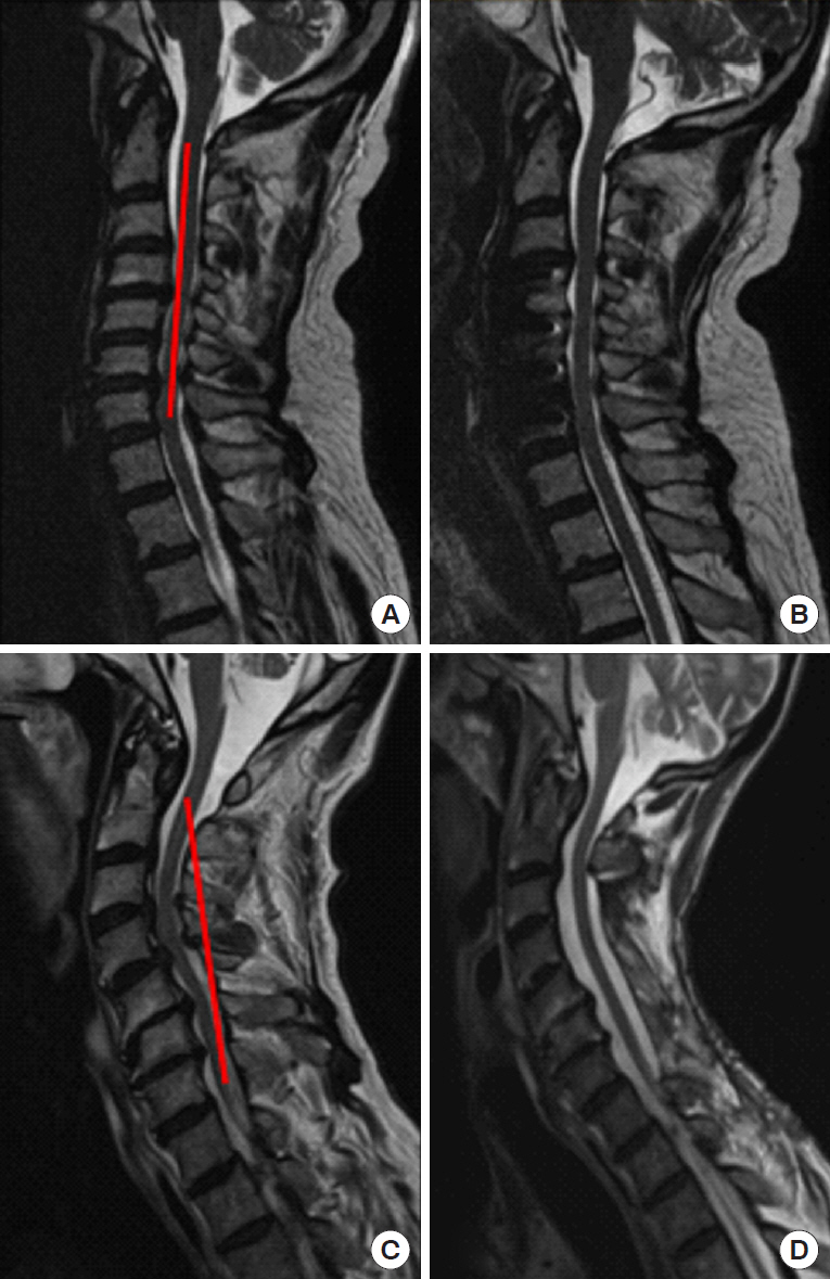
Preoperative consideration should be given to a number of factors, including age, comorbidities, cervical deformity, bone density, etc. that are proven to affect patient outcomes after DCM surgery [100]. Of these, the restoration of postoperative sagittal alignment is an important factor associated with postoperative outcomes [101]. The concept of the modified K-line and the minimum interval distance (at least 4 mm from the K-line to the anterior compressive elements) needs to be applied to every case, as failure to properly address cervical kyphosis is associated with higher risk of postoperative residual compression [100,102,103]. If the minimum interval distance is <4 mm, then an anterior approach or combined anterior/posterior approach should be strongly considered (Fig. 1), and is associated with producing better postoperative outcomes compared to posterior alone [104].
ACDF is the well-established mainstay to treat focal spondylitic cervical disease. With a predictable side effect profile and ability to treat multilevel disc disease, it has become a popular choice for day surgery intervention [105,106]. ACCF offers the ability to provide ventral decompression of retrovertebral disease or correction of kyphotic deformity with a cage or allograft construct to achieve fusion, through the same anterior approach and with a similar side effect profile [107,108]. Although no direct comparison or prospective trial exists, systematic reviews have shown multilevel ACDF is favorable above multilevel ACCF, in terms of outcome measures and sagittal alignment [109]. Hybrid constructs, using a combination of ACDF and ACCF, have also emerged as a useful tool and may be superior over a long-segment ACCF based on retrospective evidence [109-111]. Another emerging concept is the oblique corpectomy without fusion, aiming to decompress the ventral cord whilst maintaining more than 50% of the vertebral body [112]. This can be achieved using conventional ACCF techniques from a lateral approach, and advocates suggest the absence of instrumentation reduces adjacent segment degeneration but produces similar neurological outcomes compared to ACCF [113,114]. At present, however, direct comparisons between conventional ACCF and oblique corpectomy have not been performed.
For the posterior approach, laminectomy remains an excellent option for long-segment decompression, now most often combined with laminectomy and fusion (LF) to avoid the unacceptably high postlaminectomy kyphosis rates that emerged in the 1970s/1980s [115]. It is associated with a high fusion rate (>98%), with a revision rate of only 1%, and complication rate of ~9% [116]. For more focal disease amenable to the posterior approach, techniques such as ‘split’ or ‘skip’ laminectomies have become popular as a method to achieve decompression with posterior ligament and muscle attachment preservation [117,118]. These can be applied in increasingly frail patients, with similar outcomes reported compared to standard laminectomy in the limited literature that is available [118]. Laminoplasty (the technique by which the lamina is removed, ligamental decompression achieved and then the lamina ‘island’ is replaced or fused in position) was originally seen as a solution to prevent postlaminectomy kyphosis whilst avoiding the need for instrumented fusion, and remains a popular option worldwide, particularly for specific indications such as OPLL [115,119]. LF and laminoplasty have both been proven to produce neurological improvement in functional and quality of life outcome measures (up to 2 years after surgery), but comparison between the 2 techniques is difficult. A multicenter, prospective observational study comparing 166 LF patients to 100 laminoplasty showed similar patient outcomes and rates of complications, but with shorter hospital stay in the LF group [120]. Other comparisons, from systematic reviews, have favored LF to preserve cervical lordosis and to reduce neck pain, but have not found a difference in functional or quality of life metrics [87,121]. The improved lordosis with LF, however, needs to be balanced against the increased cost compared to laminoplasty, but ultimately the surgeon’s experience with either technique should be the leading discriminator [122,123].

3. Cervical Disc Arthroplasty

Cervical artificial discs (or arthroplasty) aim to preserve motion across operated segments in an effort to reduce the incidence of adjacent level disease, but have limited indications in the treatment of DCM. For soft discs and radiculopathy symptoms, it is gathering an increasing body of evidence, with increasingly long follow-up periods [124]. However, this has to be balanced against a revision rate as high as 7.7% (compared to the 2% in ACDF), revision surgeries that have increased morbidity and cost, with complications such as heterotopic ossification occurring in as high as 47% of patients [118,125-128]. These reasons, and the limitations of using CAD for more than 2 levels, have meant that there is currently only a very limited role for CAD in the treatment of DCM [100]. In addition, many surgeons believe that removing the motion across a diseased or spondilytic segment is a key component of the effectiveness of surgery and CAD is therefore contrary to this paradigm. A relatively new addition is the promotion of ‘hybrid’ constructs where CAD is used in combination with ACDF, or as an adjunct to a previous ACDF [128]. Proponents suggest that different cervical levels are subject to different mechanical stressors, and that Hybrid constructs can be used to reflect this heterogeneity between levels, however, there is a paucity of evidence to suggest Hybrid surgery is equivalent or produces different outcomes compared to CAD or ACDF.

4. Stereotactic Navigation, Robotics, and Minimally Invasive Techniques

Despite the abundance of new technology in the application of robotics, minimally invasive surgery (MIS) and stereotactic navigation in spine surgery, very little has been published with regards to improving outcomes in DCM surgery. Stereotactic navigation has been used to improve the accuracy (and therefore safety) of both cervical pedicle and lateral mass screws, in addition to aiding anterior decompression in complex craniocervical junction cases [129-131]. Robotic-assisted devices utilize stereotactic navigation to aid with pedicle screw placement, but despite their increasing popularity in North America, they currently have no role in DCM surgery [132]. The use of minimally invasive or endoscopic techniques for posterior foraminotomy have been well described, but recent reports and case series have illustrated the use of these techniques to achieve single or 2-level posterior decompression [133,134]. Tubular retractors have also been described in the application of MIS ACDF surgery, which allows for a smaller incision, less traction and greater protection from iatrogenic injury on the prevertebral soft tissues [135]. This does however come at a cost of a restricted working space and inability to use an anterior plate. Similar techniques have also been described to produce ‘tunnel’ corpectomies, however, the exact benefits of these techniques over conventional methods remain to be proven [136].

CONCLUSION AND FUTURE RESEARCH

The pathophysiology of DCM is diverse, and the range of available nonoperative and operative interventions are a testament to that fact. A large body of evidence has accumulated in recent years to demonstrate the safety and efficacy of surgical decompression in DCM, with significant gains in the functional and quality of life outcomes measured. Despite these gains, significant knowledge gaps still exist that should become the focus of future research. The current clinical assessment tools, such as the mJOA, contain a number of subjective elements and are therefore subject to interobserver discrepancies. There is a real and urgent need to develop a more objective tool to assess the severity of DCM, and the use of specialized ancillary testing (such as the GRASSP-M tool) together with quantitative imaging assessments may suit this purpose. In a similar vein, patients with ‘mild’ DCM (mJOA 15–17) often pose difficult clinical conundrums. Whereas the use of surgery is clear in moderate-severe disease, the natural history of mild DCM (or those with asymptomatic cord compression on imaging) is much more difficult to predict and therefore this cohort has become the target of recent prospective observational studies. Further still, evidence for the safe use of physiotherapy treatments and continued exercise (or elite athletic activity) in the context of mild DCM needs to be clarified. A collaborative, global effort to decide the future research priorities in DCM is currently underway [137].

CONFLICT OF INTEREST

The authors have nothing to disclose.

ACKNOWLEDGEMENTS

MGF wishes to acknowledge the Gerry and Tootsie Halbert Chair in Neural Repair and Regeneration.

Fig. 1.
T2 sagittal magnetic resonance imaging of 2 patients pre- and postoperative surgical decompression for degenerative cervical myelopathy, with the additional of the modified K-line in red. Panel A demonstrates an example of loss of the normal cervical lordosis, with the anterior compressive elements <4 mm from the modified K-line that was successfully treated with a multilevel anterior approach (B). Panel C demonstrates an example where the modified K-line does not abut the anterior elements, that was amenable to the posterior cervical approach (D).
ns-1938314-157f1.jpg

REFERENCES

1. Nurick S. The pathogenesis of the spinal cord disorder associated with cervical spondylosis. Brain 1972 95:87-100.
crossref pmid pdf
2. Karadimas SK, Erwin WM, Ely CG, et al. Pathophysiology and natural history of cervical spondylotic myelopathy. Spine (Phila Pa 1976) 2013 38(22 Suppl 1):S21-36.
crossref pmid
3. Nouri A, Tetreault L, Singh A, et al. Degenerative cervical myelopathy: epidemiology, genetics, and pathogenesis. Spine (Phila Pa 1976) 2015 40:E675-93.
crossref pmid
4. Institute of Medicine Committee on Comparative Effectiveness Research Prioritization. Initial National Priorities for Comparative Effectiveness Research. Washington, DC: National Academy of Medicine; 2009.

5. Karadimas SK, Gatzounis G, Fehlings MG. Pathobiology of cervical spondylotic myelopathy. Eur Spine J 2015 24 Suppl 2:132-8.
crossref pmid pdf
6. Klironomos G, Karadimas S, Mavrakis A, et al. New experimental rabbit animal model for cervical spondylotic myelopathy. Spinal Cord 2011 49:1097-102.
crossref pmid pdf
7. Karadimas SK, Moon ES, Yu WR, et al. A novel experimental model of cervical spondylotic myelopathy (CSM) to facilitate translational research. Neurobiol Dis 2013 54:43-58.
crossref pmid
8. Brain WR, Knight GC, Bull JW. Discussion of rupture of the intervertebral disc in the cervical region. Proc R Soc Med 1948 41:509-16.
pmid pmc
9. Breig A, Turnbull I, Hassler O. Effects of mechanical stresses on the spinal cord in cervical spondylosis. A study on fresh cadaver material. J Neurosurg 1966 25:45-56.
crossref pmid
10. Taylor AR. Mechanism and treatment of spinal-cord disorders associated with cervical spondylosis. Lancet 1953 1:717-20.
crossref pmid
11. Kalsi-Ryan S, Karadimas SK, Fehlings MG. Cervical spondylotic myelopathy: the clinical phenomenon and the current pathobiology of an increasingly prevalent and devastating disorder. Neuroscientist 2013 19:409-21.
crossref pmid
12. Karadimas SK, Klironomos G, Papachristou DJ, et al. Immunohistochemical profile of NF-κB/p50, NF-κB/p65, MMP-9, MMP-2, and u-PA in experimental cervical spondylotic myelopathy. Spine (Phila Pa 1976) 2013 38:4-10.
crossref pmid
13. Yu WR, Liu T, Kiehl TR, et al. Human neuropathological and animal model evidence supporting a role for Fas-mediated apoptosis and inflammation in cervical spondylotic myelopathy. Brain 2011 134(Pt 5):1277-92.
crossref pmid pdf
14. Hirai T, Uchida K, Nakajima H, et al. The prevalence and phenotype of activated microglia/macrophages within the spinal cord of the hyperostotic mouse (twy/twy) changes in response to chronic progressive spinal cord compression: implications for human cervical compressive myelopathy. PLoS One 2013 8:e64528.
crossref pmid pmc
15. Yu WR, Baptiste DC, Liu T, et al. Molecular mechanisms of spinal cord dysfunction and cell death in the spinal hyperostotic mouse: implications for the pathophysiology of human cervical spondylotic myelopathy. Neurobiol Dis 2009 33:149-63.
crossref pmid
16. Karadimas SK, Gialeli CH, Klironomos G, et al. The role of oligodendrocytes in the molecular pathobiology and potential molecular treatment of cervical spondylotic myelopathy. Curr Med Chem 2010 17:1048-58.
crossref pmid
17. Inukai T, Uchida K, Nakajima H, et al. Tumor necrosis factor-alpha and its receptors contribute to apoptosis of oligodendrocytes in the spinal cord of spinal hyperostotic mouse (twy/twy) sustaining chronic mechanical compression. Spine (Phila Pa 1976) 2009 34:2848-57.
crossref pmid
18. Takenouchi T, Setoguchi T, Yone K, et al. Expression of apoptosis signal-regulating kinase 1 in mouse spinal cord under chronic mechanical compression: possible involvement of the stress-activated mitogen-activated protein kinase pathways in spinal cord cell apoptosis. Spine (Phila Pa 1976) 2008 33:1943-50.
crossref pmid
19. Agrawal SK, Fehlings MG. Mechanisms of secondary injury to spinal cord axons in vitro: role of Na+, Na(+)-K(+)-ATPase, the Na(+)-H+ exchanger, and the Na(+)-Ca2+ exchanger. J Neurosci 1996 16:545-52.
crossref pmid pmc
20. Haigney MC, Miyata H, Lakatta EG, et al. Dependence of hypoxic cellular calcium loading on Na(+)-Ca2+ exchange. Circ Res 1992 71:547-57.
crossref pmid
21. Haigney MC, Lakatta EG, Stern MD, et al. Sodium channel blockade reduces hypoxic sodium loading and sodium-dependent calcium loading. Circulation 1994 90:391-9.
crossref pmid
22. Regan RF, Choi DW. Glutamate neurotoxicity in spinal cord cell culture. Neuroscience 1991 43:585-91.
crossref pmid
23. Schwartz G, Fehlings MG. Secondary injury mechanisms of spinal cord trauma: a novel therapeutic approach for the management of secondary pathophysiology with the sodium channel blocker riluzole. Prog Brain Res 2002 137:177-90.
crossref pmid
24. Kopjar B, Tetreault L, Kalsi-Ryan S, et al. Psychometric properties of the modified Japanese Orthopaedic Association scale in patients with cervical spondylotic myelopathy. Spine (Phila Pa 1976) 2015 40:E23-8.
crossref pmid
25. Furlan JC, Catharine Craven B. Psychometric analysis and critical appraisal of the original, revised, and modified versions of the Japanese Orthopaedic Association score in the assessment of patients with cervical spondylotic myelopathy. Neurosurg Focus 2016 40:E6.
crossref pmc
26. Zhou F, Zhang Y, Sun Y, et al. Assessment of the minimum clinically important difference in neurological function and quality of life after surgery in cervical spondylotic myelopathy patients: a prospective cohort study. Eur Spine J 2015 24:2918-23.
crossref pmid pdf
27. Singh A, Crockard HA. Comparison of seven different scales used to quantify severity of cervical spondylotic myelopathy and post-operative improvement. J Outcome Meas 2001-2002 5:798-818.

28. Kalsi-Ryan S, Singh A, Massicotte EM, et al. Ancillary outcome measures for assessment of individuals with cervical spondylotic myelopathy. Spine (Phila Pa 1976) 2013 38(22 Suppl 1):S111-22.
crossref pmid
29. Zhan A, Mohan S, Tarolli C, et al. Using smartphones and machine learning to quantify parkinson disease severity: the Mobile Parkinson Disease Score. JAMA Neurol 2018 75:876-80.
crossref pmid pmc
30. Stewart M, Smith S, Davies B, et al. P90 A systematic review of spinal cord serum and cerebrospinal fluid biomarkers for use in degenerative cervical myelopathy. J Neurol Neurosurg Psychiatr 2019 90:e45-6.
crossref
31. Laliberte AM. An examination of the role of MicroRNA-21 in the pathobiology of degenerative cervical myelopathy using human and animal data. Toronto: Institute of Medical Science, University of Toronto; 2018.

32. Xu C, Zhang H, Zhou W, et al. MicroRNA-10a, -210, and -563 as circulating biomarkers for ossification of the posterior longitudinal ligament. Spine J 2019 19:735-43.
crossref pmid
33. Jutzeler CR, Ulrich A, Huber B, et al. Improved diagnosis of cervical spondylotic myelopathy with contact heat evoked potentials. J Neurotrauma 2017 34:2045-53.
crossref pmid
34. Nouri A, Martin AR, Kato S, et al. The relationship between MRI signal intensity changes, clinical presentation, and surgical outcome in degenerative cervical myelopathy: analysis of a global cohort. Spine (Phila Pa 1976) 2017 42:1851-8.
crossref pmid
35. Martin AR, Aleksanderek I, Cohen-Adad J, et al. Translating state-of-the-art spinal cord MRI techniques to clinical use: a systematic review of clinical studies utilizing DTI, MT, MWF, MRS, and fMRI. Neuroimage Clin 2015 10:192-238.
crossref pmid pmc
36. Martin AR, De Leener B, Cohen-Adad J, et al. Can microstructural MRI detect subclinical tissue injury in subjects with asymptomatic cervical spinal cord compression? A prospective cohort study. BMJ Open 2018 8:e019809.
crossref pmid pmc
37. Martin AR, De Leener B, Cohen-Adad J, et al. Clinically feasible microstructural MRI to quantify cervical spinal cord tissue injury using DTI, MT, and T2*-weighted imaging: assessment of normative data and reliability. AJNR Am J Neuroradiol 2017 38:1257-65.
crossref pmid pmc
38. Yoo WK, Kim TH, Hai DM, et al. Correlation of magnetic resonance diffusion tensor imaging and clinical findings of cervical myelopathy. Spine J 2013 13:867-76.
crossref pmid
39. Grabher P, Mohammadi S, David G, et al. Neurodegeneration in the spinal ventral horn prior to motor impairment in cervical spondylotic myelopathy. J Neurotrauma 2017 34:2329-34.
crossref pmid
40. Martin AR, De Leener B, Cohen-Adad J, et al. A novel MRI biomarker of spinal cord white matter injury: T2*-weighted white matter to gray matter signal intensity ratio. AJNR Am J Neuroradiol 2017 38:1266-73.
crossref pmid pmc
41. De Leener B, Lévy S, Dupont SM, et al. CT: spinal cord toolbox, an open-source software for processing spinal cord MRI data. Neuroimage 2017 145(Pt A):24-43.
crossref pmid
42. Merali ZG, Witiw CD, Badhiwala JH, et al. Using a machine learning approach to predict outcome after surgery for degenerative cervical myelopathy. PLoS One 2019 14:e0215133.
crossref pmid pmc
43. Badhiwala JH, Hachem LD, Merali Z, et al. Predicting outcomes after surgical decompression for mild degenerative cervical myelopathy: moving beyond the mJOA to identify surgical candidates. Neurosurgery 2019 Jun 21 [Epub]. pii: nyz160. https://doi.org/10.1093/neuros/nyz160.
crossref pdf
44. Badhiwala JH, Witiw CD, Nassiri F, et al. Efficacy and safety of surgery for mild degenerative cervical myelopathy: results of the AOSpine North America and International Prospective Multicenter Studies. Neurosurgery 2019 84:890-7.
crossref pmid pdf
45. Kadaňka Z, Bednařík J, Novotný O, et al. Cervical spondylotic myelopathy: conservative versus surgical treatment after 10 years. Eur Spine J 2011 20:1533-8.
crossref pmid pmc pdf
46. Rhee J, Tetreault LA, Chapman JR, et al. Nonoperative versus operative management for the treatment degenerative cervical myelopathy: an updated systematic review. Global Spine J 2017 7(3 Suppl):35S-41S.
crossref pmid pmc
47. Rhee JM, Shamji MF, Erwin WM, et al. Nonoperative management of cervical myelopathy: a systematic review. Spine (Phila Pa 1976) 2013 38(22 Suppl 1):S55-67.
crossref pmid
48. Miller RG, Mitchell JD, Lyon M, et al. Riluzole for amyotrophic lateral sclerosis (ALS)/motor neuron disease (MND). Cochrane database of systematic reviews. Hoboken (NJ): John Wiley & Sons, Ltd.; 2002.

49. Mizoule J, Meldrum B, Mazadier M, et al. 2-Amino-6-trifluoromethoxy benzothiazole, a possible antagonist of excitatory amino acid neurotransmission--I. Anticonvulsant properties. Neuropharmacology 1985 24:767-73.
crossref pmid
50. Fehlings MG, Wilson JR, Karadimas SK, et al. Clinical evaluation of a neuroprotective drug in patients with cervical spondylotic myelopathy undergoing surgical treatment: design and rationale for the CSM-Protect trial. Spine (Phila Pa 1976) 2013 38(22 Suppl 1):S68-75.
crossref pmid
51. Karadimas SK, Laliberte AM, Tetreault L, et al. Riluzole blocks perioperative ischemia-reperfusion injury and enhances postdecompression outcomes in cervical spondylotic myelopathy. Sci Transl Med 2015 7:316ra194.
crossref pmid
52. Satkunendrarajah K, Nassiri F, Karadimas SK, et al. Riluzole promotes motor and respiratory recovery associated with enhanced neuronal survival and function following high cervical spinal hemisection. Exp Neurol 2016 276:59-71.
crossref pmid
53. Michael Fehlings BK, Badhiwala J, Ahn H, et al. The safety and efficacy of riluzole in enhancing clinical outcomes in patients undergoing surgery for cervical spondylotic myelopathy: results of the CSM-Protect double-blinded, multicentre randomized controlled trial in 300 patients. Can J Surg 2019 62(4 Suppl 1):S46.

54. Vidal PM, Karadimas SK, Ulndreaj A, et al. Delayed decompression exacerbates ischemia-reperfusion injury in cervical compressive myelopathy. JCI Insight 2017 Jun 2 2(11):pii: 92512. https://doi.org/10.1172/jci.insight.92512.
crossref
55. Li XQ, Lv HW, Tan WF, et al. Role of the TLR4 pathway in blood-spinal cord barrier dysfunction during the bimodal stage after ischemia/reperfusion injury in rats. J Neuroinflammation 2014 11:62.
crossref pmid pmc
56. Li XQ, Wang J, Fang B, et al. Intrathecal antagonism of microglial TLR4 reduces inflammatory damage to blood-spinal cord barrier following ischemia/reperfusion injury in rats. Mol Brain 2014 7:28.
crossref pmid pmc
57. Zhang N, Komine-Kobayashi M, Tanaka R, et al. Edaravone reduces early accumulation of oxidative products and sequential inflammatory responses after transient focal ischemia in mice brain. Stroke 2005 36:2220-5.
crossref pmid
58. Vidal PM, Ulndreaj A, Badner A, et al. Methylprednisolone treatment enhances early recovery following surgical decompression for degenerative cervical myelopathy without compromise to the systemic immune system. J Neuroinflammation 2018 15:222.
crossref pmid pmc pdf
59. Bracken MB, Shepard MJ, Collins WF, et al. A randomized, controlled trial of methylprednisolone or naloxone in the treatment of acute spinal-cord injury. Results of the Second National Acute Spinal Cord Injury Study. N Engl J Med 1990 322:1405-11.
crossref pmid
60. Evaniew N, Belley-Côté EP, Fallah N, et al. Methylprednisolone for the treatment of patients with acute spinal cord injuries: a systematic review and meta-analysis. J Neurotrauma 2016 33:468-81.
crossref pmid pmc
61. Evaniew N, Noonan VK, Fallah N, et al. Methylprednisolone for the treatment of patients with acute spinal cord injuries: a propensity score-matched cohort study from a Canadian multi-center spinal cord injury registry. J Neurotrauma 2015 32:1674-83.
crossref pmid pmc
62. Jeyamohan SB, Kenning TJ, Petronis KA, et al. Effect of steroid use in anterior cervical discectomy and fusion: a randomized controlled trial. J Neurosurg Spine 2015 23:137-43.
crossref pmid
63. Aljabi Y, El-Shawarby A, Cawley DT, et al. Effect of epidural methylprednisolone on post-operative pain and length of hospital stay in patients undergoing lumbar microdiscectomy. Surgeon 2015 13:245-9.
crossref pmid
64. Ghasemi M, Masaeli A, Rezvani M, et al. Oral prednisolone in the treatment of cervical radiculopathy: a randomized placebo controlled trial. J Res Med Sci 2013 18(Suppl 1):S43-6.
pmid pmc
65. Meyer F, Börm W, Thomé C. Degenerative cervical spinal stenosis: current strategies in diagnosis and treatment. Dtsch Arztebl Int 2008 105:366-72.
crossref pmid pmc
66. Bergknut N, Rutges JP, Kranenburg HJ, et al. The dog as an animal model for intervertebral disc degeneration? Spine (Phila Pa 1976) 2012 37:351-8.
crossref pmid
67. Erwin WM, DeSouza L, Funabashi M, et al. The biological basis of degenerative disc disease: proteomic and biomechanical analysis of the canine intervertebral disc. Arthritis Res Ther 2015 17:240.
crossref pmid pmc
68. Imai Y, Okuma M, An HS, et al. Restoration of disc height loss by recombinant human osteogenic protein-1 injection into intervertebral discs undergoing degeneration induced by an intradiscal injection of chondroitinase ABC. Spine (Phila Pa 1976) 2007 32:1197-205.
crossref pmid
69. Masuda K, Imai Y, Okuma M, et al. Osteogenic protein-1 injection into a degenerated disc induces the restoration of disc height and structural changes in the rabbit anular puncture model. Spine (Phila Pa 1976) 2006 31:742-54.
crossref pmid
70. Walsh AJ, Bradford DS, Lotz JC. In vivo growth factor treatment of degenerated intervertebral discs. Spine (Phila Pa 1976) 2004 29:156-63.
crossref pmid
71. Wei A, Williams LA, Bhargav D, et al. BMP13 prevents the effects of annular injury in an ovine model. Int J Biol Sci 2009 5:388-96.
crossref pmid pmc
72. Leckie SK, Bechara BP, Hartman RA, et al. Injection of AAV2-BMP2 and AAV2-TIMP1 into the nucleus pulposus slows the course of intervertebral disc degeneration in an in vivo rabbit model. Spine J 2012 12:7-20.
crossref pmid
73. Nishida K, Doita M, Takada T, et al. Sustained transgene expression in intervertebral disc cells in vivo mediated by microbubble-enhanced ultrasound gene therapy. Spine (Phila Pa 1976) 2006 31:1415-9.
crossref pmid
74. Paul R, Haydon RC, Cheng H, et al. Potential use of Sox9 gene therapy for intervertebral degenerative disc disease. Spine (Phila Pa 1976) 2003 28:755-63.
crossref pmid pmc
75. Seki S, Asanuma-Abe Y, Masuda K, et al. Effect of small interference RNA (siRNA) for ADAMTS5 on intervertebral disc degeneration in the rabbit anular needle-puncture model. Arthritis Res Ther 2009 11:R166.
crossref pmid pmc
76. Zhang YH, Zhao CQ, Jiang LS, et al. Lentiviral shRNA silencing of CHOP inhibits apoptosis induced by cyclic stretch in rat annular cells and attenuates disc degeneration in the rats. Apoptosis 2011 16:594-605.
crossref pmid pdf
77. Bae WC, Masuda K. Emerging technologies for molecular therapy for intervertebral disk degeneration. Orthop Clin North Am 2011 42:585. -601. ix.
crossref pmid pmc
78. Dowdell J, Erwin M, Choma T, et al. Intervertebral disk degeneration and repair. Neurosurgery 2017 80(3S):S46-54.
crossref pmid pmc pdf
79. Urits I, Capuco A, Sharma M, et al. Stem cell therapies for treatment of discogenic low back pain: a comprehensive review. Curr Pain Headache Rep 2019 23:65.
crossref pmid pdf
80. Wang Z, Perez-Terzic CM, Smith J, et al. Efficacy of intervertebral disc regeneration with stem cells - a systematic review and meta-analysis of animal controlled trials. Gene 2015 564:1-8.
crossref pmid
81. Meisel HJ, Ganey T, Hutton WC, et al. Clinical experience in cell-based therapeutics: intervention and outcome. Eur Spine J 2006 15 Suppl 3:S397-405.
crossref pmid pdf
82. Meisel HJ, Siodla V, Ganey T, et al. Clinical experience in cell-based therapeutics: disc chondrocyte transplantation A treatment for degenerated or damaged intervertebral disc. Biomol Eng 2007 24:5-21.
crossref pmid
83. Orozco L, Soler R, Morera C, et al. Intervertebral disc repair by autologous mesenchymal bone marrow cells: a pilot study. Transplantation 2011 92:822-8.
crossref pmid
84. Yoshikawa T, Ueda Y, Miyazaki K, et al. Disc regeneration therapy using marrow mesenchymal cell transplantation: a report of two case studies. Spine (Phila Pa 1976) 2010 35:E475-80.
crossref pmid
85. Kurd MF, Kreitz T, Schroeder G, et al. The role of multimodal analgesia in spine surgery. J Am Acad Orthop Surg 2017 25:260-8.
crossref pmid
86. Devin CJ, McGirt MJ. Best evidence in multimodal pain management in spine surgery and means of assessing postoperative pain and functional outcomes. J Clin Neurosci 2015 22:930-8.
crossref pmid
87. Lee CH, Lee J, Kang JD, et al. Laminoplasty versus laminectomy and fusion for multilevel cervical myelopathy: a meta-analysis of clinical and radiological outcomes. J Neurosurg Spine 2015 22:589-95.
crossref pmid
88. Woolf CJ, Chong MS. Preemptive analgesia--treating postoperative pain by preventing the establishment of central sensitization. Anesth Analg 1993 77:362-79.
crossref pmid
89. Chang WK, Wu HL, Yang CS, et al. Effect on pain relief and inflammatory response following addition of tenoxicam to intravenous patient-controlled morphine analgesia: a double-blind, randomized, controlled study in patients undergoing spine fusion surgery. Pain Med 2013 14:736-48.
crossref pmid pdf
90. Dodwell ER, Latorre JG, Parisini E, et al. NSAID exposure and risk of nonunion: a meta-analysis of case-control and cohort studies. Calcif Tissue Int 2010 87:193-202.
crossref pmid pdf
91. Jirarattanaphochai K, Jung S. Nonsteroidal antiinflammatory drugs for postoperative pain management after lumbar spine surgery: a meta-analysis of randomized controlled trials. J Neurosurg Spine 2008 9:22-31.
crossref pmid
92. Li Q, Zhang Z, Cai Z. High-dose ketorolac affects adult spinal fusion: a meta-analysis of the effect of perioperative nonsteroidal anti-inflammatory drugs on spinal fusion. Spine (Phila Pa 1976) 2011 36:E461-8.
crossref pmid
93. Backonja M, Beydoun A, Edwards KR, et al. Gabapentin for the symptomatic treatment of painful neuropathy in patients with diabetes mellitus: a randomized controlled trial. JAMA 1998 280:1831-6.
crossref pmid
94. Lesser H, Sharma U, LaMoreaux L, et al. Pregabalin relieves symptoms of painful diabetic neuropathy: a randomized controlled trial. Neurology 2004 63:2104-10.
crossref pmid
95. Lo YL, Cheong PW, George JM, et al. Pregabalin and Radicular Pain Study (PARPS) for cervical spondylosis in a multiracial Asian population. J Clin Med Res 2014 6:66-71.
crossref pmid
96. Fehlings MG, Barry S, Kopjar B, et al. Anterior versus posterior surgical approaches to treat cervical spondylotic myelopathy: outcomes of the prospective multicenter AOSpine North America CSM study in 264 patients. Spine (Phila Pa 1976) 2013 38:2247-52.
crossref pmid
97. Asher AL, Devin CJ, Kerezoudis P, et al. Comparison of outcomes following anterior vs posterior fusion surgery for patients with degenerative cervical myelopathy: an analysis from quality outcomes database. Neurosurgery 2019 84:919-26.
crossref pmid pdf
98. Kato S, Nouri A, Wu D, et al. Comparison of anterior and posterior surgery for degenerative cervical myelopathy: an MRI-based propensity-score-matched analysis using data from the prospective multicenter AOSpine CSM North America and International Studies. J Bone Joint Surg Am 2017 99:1013-21.
crossref pmid
99. Ghogawala Z, Benzel EC, Heary RF, et al. Cervical spondylotic myelopathy surgical trial: randomized, controlled trial design and rationale. Neurosurgery 2014 75:334-46.
crossref pmid pmc pdf
100. Wilson J, Jiang F, Fehlings M. Clinical predictors of complications and outcomes in degenerative cervical myeloradiculopathy. Indian Spine J 2019 2:59-67.
crossref
101. Uchida K, Nakajima H, Sato R, et al. Cervical spondylotic myelopathy associated with kyphosis or sagittal sigmoid alignment: outcome after anterior or posterior decompression. J Neurosurg Spine 2009 11:521-8.
crossref pmid
102. Taniyama T, Hirai T, Yamada T, et al. Modified K-line in magnetic resonance imaging predicts insufficient decompression of cervical laminoplasty. Spine (Phila Pa 1976) 2013 38:496-501.
crossref pmid
103. Taniyama T, Hirai T, Yoshii T, et al. Modified K-line in magnetic resonance imaging predicts clinical outcome in patients with nonlordotic alignment after laminoplasty for cervical spondylotic myelopathy. Spine (Phila Pa 1976) 2014 39:E1261-8.
crossref pmid
104. Shamji MF, Mohanty C, Massicotte EM, et al. The association of cervical spine alignment with neurologic recovery in a prospective cohort of patients with surgical myelopathy: analysis of a series of 124 cases. World Neurosurg 2016 86:112-9.
crossref pmid
105. Ban D, Liu Y, Cao T, et al. Safety of outpatient anterior cervical discectomy and fusion: a systematic review and meta-analysis. Eur J Med Res 2016 21:34.
crossref pmid pmc
106. McClelland S 3rd, Oren JH, Protopsaltis TS, et al. Outpatient anterior cervical discectomy and fusion: a meta-analysis. J Clin Neurosci 2016 34:166-8.
crossref pmid
107. Douglas AF, Cooper PR. Cervical corpectomy and strut grafting. Neurosurgery 2007 60(1 Supp1 1):S137-42.
crossref pmid pdf
108. Ghogawala Z. Anterior cervical option to manage degenerative cervical myelopathy. Neurosurg Clin N Am 2018 29:83-9.
crossref pmid
109. Shamji MF, Massicotte EM, Traynelis VC, et al. Comparison of anterior surgical options for the treatment of multilevel cervical spondylotic myelopathy: a systematic review. Spine (Phila Pa 1976) 2013 38(22 Suppl 1):S195-209.
crossref pmid
110. Ashkenazi E, Smorgick Y, Rand N, et al. Anterior decompression combined with corpectomies and discectomies in the management of multilevel cervical myelopathy: a hybrid decompression and fixation technique. J Neurosurg Spine 2005 3:205-9.
crossref pmid
111. Liu Y, Yu KY, Hu JH. Hybrid decompression technique and two-level corpectomy are effective treatments for three-level cervical spondylotic myelopathy. J Zhejiang Univ Sci B 2009 10:696-701.
crossref pmid pmc pdf
112. George B, Gauthier N, Lot G. Multisegmental cervical spondylotic myelopathy and radiculopathy treated by multilevel oblique corpectomies without fusion. Neurosurgery 1999 44:81-90.
crossref pmid pdf
113. Chibbaro S, Benvenuti L, Carnesecchi S, et al. Anterior cervical corpectomy for cervical spondylotic myelopathy: experience and surgical results in a series of 70 consecutive patients. J Clin Neurosci 2006 13:233-8.
crossref pmid
114. Kiris T, Kilinçer C. Cervical spondylotic myelopathy treated by oblique corpectomy: a prospective study. Neurosurgery 2008 62:674-82.
crossref pmid pdf
115. Abduljabbar FH, Teles AR, Bokhari R, et al. Laminectomy with or without fusion to manage degenerative cervical myelopathy. Neurosurg Clin N Am 2018 29:91-105.
crossref pmid
116. Youssef JA, Heiner AD, Montgomery JR, et al. Outcomes of posterior cervical fusion and decompression: a systematic review and meta-analysis. Spine J 2019 May 7 [Epub]. pii: S1529-9430(19)30166-4. https://doi.org/10.1016/j.spinee.2019.04.019.
crossref
117. Luo W, Li Y, Zhao J, et al. Skip Laminectomy compared with laminoplasty for cervical compressive myelopathy: a systematic review and meta-analysis. World Neurosurg 2018 120:296-301.
crossref pmid
118. Traynelis VC, Arnold PM, Fourney DR, et al. Alternative procedures for the treatment of cervical spondylotic myelopathy: arthroplasty, oblique corpectomy, skip laminectomy: evaluation of comparative effectiveness and safety. Spine (Phila Pa 1976) 2013 38(22 Suppl 1):S210-31.
crossref pmid
119. Wilson JR, Tetreault LA, Kim J, et al. State of the art in degenerative cervical myelopathy: an update on current clinical evidence. Neurosurgery 2017 80(3S):S33-45.
crossref pmid pdf
120. Fehlings MG, Santaguida C, Tetreault L, et al. Laminectomy and fusion versus laminoplasty for the treatment of degenerative cervical myelopathy: results from the AOSpine North America and International prospective multicenter studies. Spine J 2017 17:102-8.
crossref pmid
121. Yoon ST, Hashimoto RE, Raich A, et al. Outcomes after laminoplasty compared with laminectomy and fusion in patients with cervical myelopathy: a systematic review. Spine (Phila Pa 1976) 2013 38(22 Suppl 1):S183-94.
crossref pmid
122. Blizzard DJ, Caputo AM, Sheets CZ, et al. Laminoplasty versus laminectomy with fusion for the treatment of spondylotic cervical myelopathy: short-term follow-up. Eur Spine J 2017 26:85-93.
crossref pdf
123. Lau D, Winkler EA, Than KD, et al. Laminoplasty versus laminectomy with posterior spinal fusion for multilevel cervical spondylotic myelopathy: influence of cervical alignment on outcomes. J Neurosurg Spine 2017 27:508-17.
crossref pmid
124. Latka D, Kozlowska K, Miekisiak G, et al. Safety and efficacy of cervical disc arthroplasty in preventing the adjacent segment disease: a meta-analysis of mid- to long-term outcomes in prospective, randomized, controlled multicenter studies. Ther Clin Risk Manag 2019 15:531-9.
crossref pmid pmc
125. Nandyala SV, Marquez-Lara A, Fineberg SJ, et al. Comparison of revision surgeries for one- to two-level cervical TDR and ACDF from 2002 to 2011. Spine J 2014 14:2841-6.
crossref pmid
126. Fay LY, Huang WC, Wu JC, et al. Arthroplasty for cervical spondylotic myelopathy: similar results to patients with only radiculopathy at 3 years’ follow-up. J Neurosurg Spine 2014 21:400-10.
crossref pmid
127. Zheng B, Hao D, Guo H, et al. ACDF vs TDR for patients with cervical spondylosis - an 8 year follow up study. BMC Surg 2017 17:113.
crossref pmid pmc pdf
128. Laratta JL, Shillingford JN, Saifi C, et al. Cervical disc arthroplasty: a comprehensive review of single-level, multilevel, and hybrid procedures. Global Spine J 2018 8:78-83.
crossref pmid
129. Arab A, Alkherayf F, Sachs A, et al. Use of 3D navigation in subaxial cervical spine lateral mass screw insertion. J Neurol Surg Rep 2018 79:e1-8.
crossref pmid pmc pdf
130. Shimokawa N, Takami T. Surgical safety of cervical pedicle screw placement with computer navigation system. Neurosurg Rev 2017 40:251-8.
crossref pmid pdf
131. Miyahara J, Hirao Y, Matsubayashi Y, et al. Computer tomography navigation for the transoral anterior release of a complex craniovertebral junction deformity: a report of two cases. Int J Surg Case Rep 2016 24:142-5.
crossref pmid pmc
132. Malham GM, Wells-Quinn T. What should my hospital buy next?-Guidelines for the acquisition and application of imaging, navigation, and robotics for spine surgery. J Spine Surg 2019 5:155-65.
crossref pmid pmc
133. Hernandez RN, Wipplinger C, Navarro-Ramirez R, et al. Ten-step minimally invasive cervical decompression via unilateral tubular laminotomy: technical note and early clinical experience. Oper Neurosurg (Hagerstown) 2019 Jun 27 [Epub]. pii: opz156. https://doi.org/10.1093/ons/opz156.
crossref pdf
134. Lin Y, Rao S, Li Y, et al. Posterior percutaneous full-endoscopic cervical laminectomy and decompression for cervical stenosis with myelopathy: a technical note. World Neurosurg 2019 Jan 12 pii: S1878-8750(19)30051-8 [Epub]. https://doi.org/10.1016/j.wneu.2018.12.180.
crossref
135. Vergara P, Timofeev I. Minimally invasive anterior cervical discectomy and fusion: a valid alternative to open techniques. Acta Neurochir (Wien) 2018 160:2467-71.
crossref pmid pdf
136. Quillo-Olvera J, Lin GX, Suen TK, et al. Anterior transcorporeal tunnel approach for cervical myelopathy guided by CT-based intraoperative spinal navigation: technical note. J Clin Neurosci 2018 48:218-23.
crossref pmid
137. Davies BM, Khan DZ, Mowforth OD, et al. RE-CODE DCM (REsearch Objectives and Common Data Elements for Degenerative Cervical Myelopathy): a consensus process to improve research efficiency in DCM, through establishment of a standardized dataset for clinical research and the definition of the research priorities. Global Spine J 2019 9(1 Suppl):65S-76S.
crossref pmid pmc


Editorial Office
CHA University, CHA School of Medicine Bundang Medical Center
59 Yatap-ro, Bundang-gu, Seongnam-si, Gyeonggi-do 13496, Korea
Tel: +82-31-780-1924  Fax: +82-31-780-5269  E-mail: support@e-neurospine.org
The Korean Spinal Neurosurgery Society
#407, Dong-A Villate 2nd Town, 350 Seocho-daero, Seocho-gu, Seoul 06631, Korea
Tel: +82-2-585-5455  Fax: +82-2-2-523-6812  E-mail: ksns1987@gmail.com
Business License No.: 209-82-62443

Copyright © The Korean Spinal Neurosurgery Society.

Developed in M2PI

Close layer
prev next