


INTRODUCTION
Vertebral column is a common site for bony metastasis in patients with systemic malignancy. But if we consider metastasis of adenocarcinoma, especially vertebral metastasis of intraductal papillary mucinous neoplasm(IPMN) is rare4,7). IPMN is a pancreatic neoplasm, characterized by production of mucinous fluid, cystic dilatation of the pancreatic ducts, and intrapapillary growth2,8,9). Mucin could be produced by metastatic spinal tumor originating from pancreatic IPMN as the primary tumor. However, no report has addressed the mucin-associated complication after spinal surgery for metastatic pancreatic cancer.
Here, we report extremely rare case of delayed large epidural mucin collection causing neurologic deficit after surgery for metastatic pancreatic cancer.
CASE REPORT
A 65-year-old man presented with abdominal discomfort and intractable upper-thoracic back pain radiating to the chest and gait disturbance. He had a history of subtotal pancreatectomy and total splenectomy due to IPMN of the pancreas at another hospital. The diameter of tumor mass was 5.2 cm, and histopathologic finding confirmed that there was invasion of peripancreatic soft tissue and lymph node metastasis. He underwent chemotherapy with gemcitabine hydrochloride as adjuvant therapy. Eight months after subtotal pancreatectomy, no specific symptoms were developed but spinal metastasis was found at T2 and T4 on a first positron emission tomography-computed tomography. Radiotherapy delivering 44 Gy in 20 fractions was applied to spinal bone metastasis and TS-1 chemotherapy was subsequently implemented. Sixteen months after pancreatectomy, weakness in lower extremities (strength 4/5) and gait disturbance were occurred and thoracic magnetic resonance imaging (MRI) showed interval aggravation of metastasis at T2 and T4 involving epidural space and posterior element (Fig. 1). We performed total laminectomy and pediculectomy T2 and T4 with subtotal resection of the tumor masses for decompression, and pedicle screw fixation at C7ŌĆōT6 for stabilization (Fig. 2). After the operation, patientŌĆÖs lower limb motor was nearly intact, and he could walk without support.
Fig.┬Ā1
Axial (A) and sagittal T2-(B) and contrast enhanced T1-weighted (C) magnetic resonance imaging shows vertebral collapse and cord compromise due to metastatic tumors of T2 and T4.
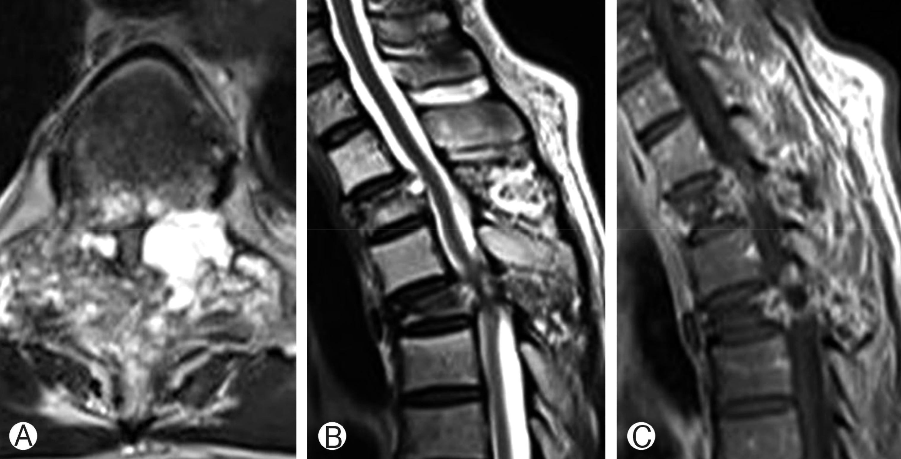
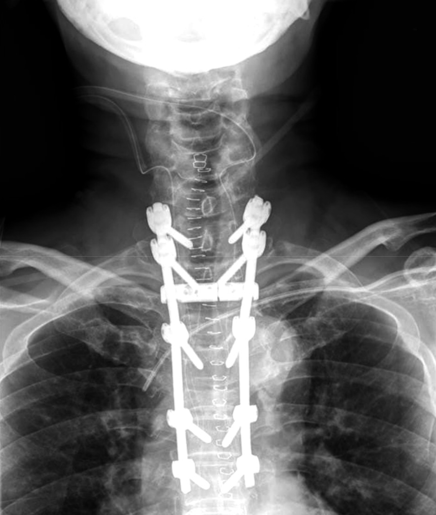
Fig.┬Ā2
Postoperative image of total laminectomy and pediculectomy T2 and T4 with subtotal resection of the tumor masses, and pedicle screw fixation at C7ŌĆōT6.
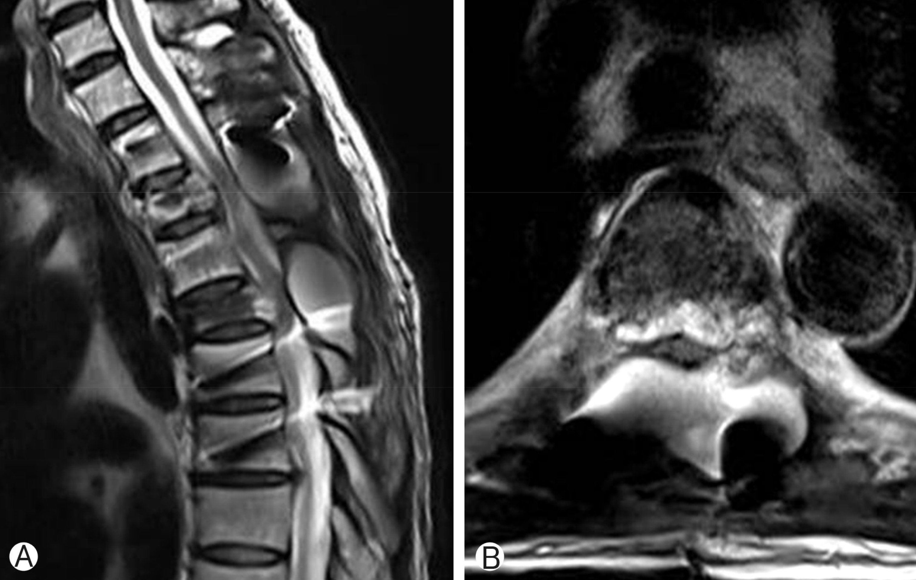
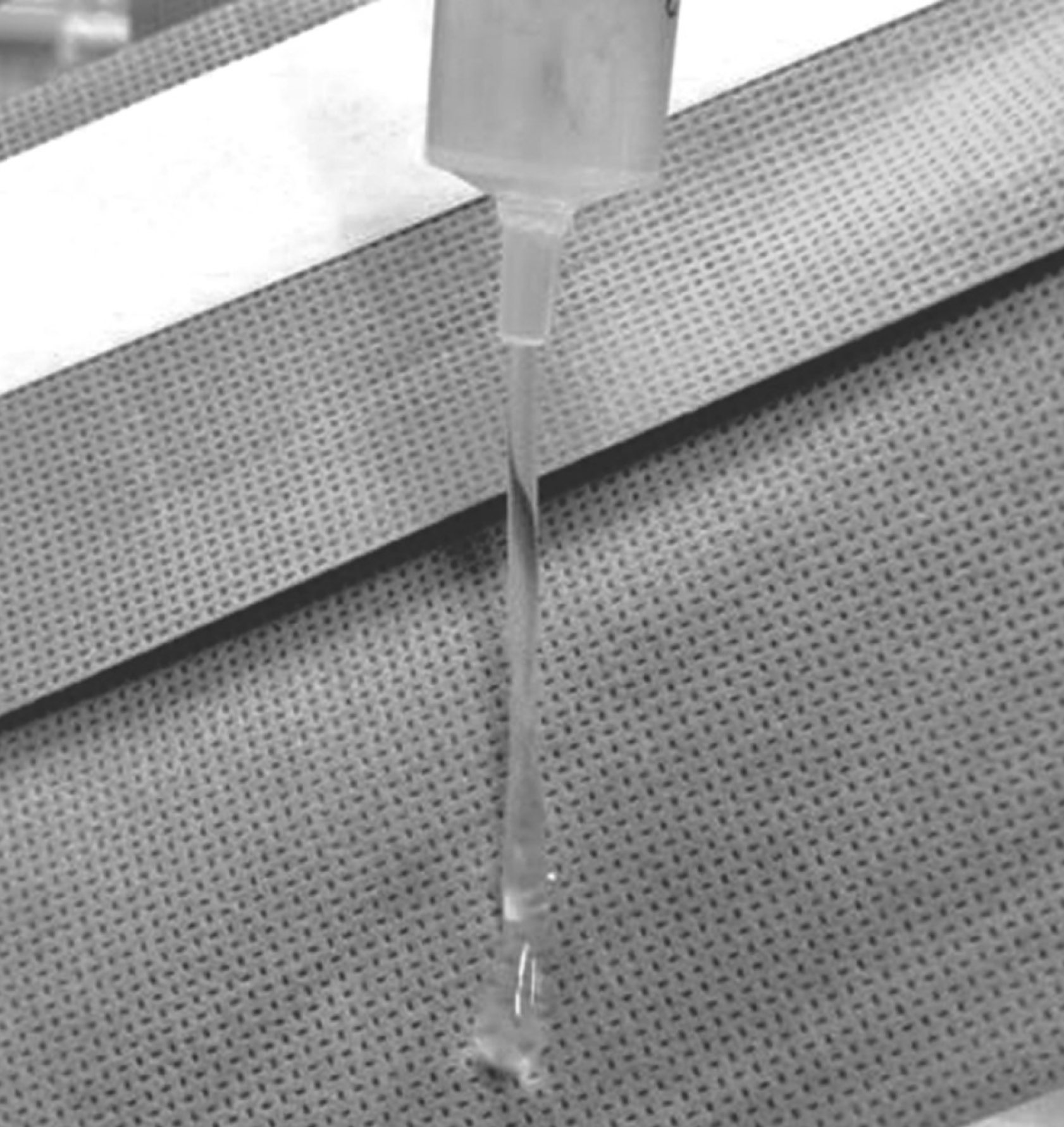
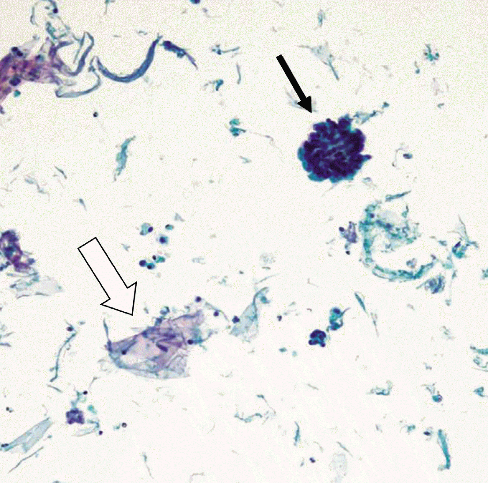
Three months after spinal surgery, motor weakness and paresthesia reappeared. MRI showed significant loculated fluid collection around the laminectomy site which resulted in compression of the spinal cord at the level of T4 (Fig. 3). We performed wound revision and large amount of fluid was expelled out. The nature of the fluid was transparent, mucoid, and sticky (Fig. 4). Total volume of removed fluid was about 150 mL. Liquid based cytology examination showed the fluid component as mucin and tumor cells (Fig. 5). After the revision, motor power of lower extremities was improved, but small amount of mucin was continually expelled through a drainage site of the skin requiring repetitive paracentesis. At that time, patient complained persistent abdominal discomfort, abdominal computed tomography revealed new appearance of carcinomatosis peritonei. Six months after first spine surgery, he expired due to deteriorated pneumonia.
DISCUSSION
In most cases, IPMN of the pancreas is a slow-growing tumor and shows excellent prognosis4). However, invasive IPMN has a high recurrence and high mortality8). In particular, lymph node metastasis is an important prognostic factor and frequent in patients with invasive IPMNs7). For those patients with invasive IPMNs, 1-, 2-year actuarial survival rates were 45%, 24% with positive lymph nodes, on the other hand 95%, 95% with negative lymph nodes6). In addition, distant metastasis may be a poor prognostic factor4). In our case, because of poor prognosis of invasive IPMN with positive lymph node metastasis, we selected subtotal removal of tumor mass as an operative method. Unlike lymph node metastasis, distant metastasis of IPMNs is rare1,7). Liver and lung have been reported as the common sites of distant metastasis7,8). In particular, vertebral metastasis of invasive IPMN is extremely rare and only one case has been reported7).
Characteristically, IPMNs of the pancreas produce excessive gelatinous mucin by ductal epithelial cells which may cause biliary obstruction, pancreatitis, and fibrosis5). The excessive mucin production of the IPMN can occasionally cause rupture of the pancreatic duct, resulting in spillage of mucinous material into the peritoneal cavity and pseudomxyoma peritonei5). Metastatic tumor of IPMNs may also produce mucin materials. However, complications associated with excessive mucin collection of metastatic IPMNs have rarely been reported, especially in vertebral metastasis. In our case, subtotal tumor debulking, laminectomy and fixation with pedicle screw insertion were performed in a routine manner3). However, remnant metastatic tumor cells continually produced mucin. Accumulated mucin at the laminectomy site compressed the spinal cord, resulting in neurologic aggravation 3 months after surgery. The current case is the first report of mucin-associated complication of metastatic IPMNs in spine.
No definite guidelines for treatment of extrapancreatic mucin have been reported. In cases of pseudomyxoma peritonei by IPMNs, initial treatment includes radical, repetitive debulking operations with complete mucus removal5). However, radical tumor resection and mucin removal may be limited in metastatic spinal tumor. Beside the radical operation, many alternative treatments have been applied including mucolytic agents, phototherapy, adjuvant systemic chemotherapy, and radiotherapy5). However, clinical course of pseudomyxoma peritonei ultimately shows an overall poor prognosis4,8). In our case, radiotherapy with concurrent chemotherapy was not effective in reducing tumor burden. Instead, repetitive paracentesis was performed after revision surgery in order to prevent spinal cord compression by accumulation of mucin.
CONCLUSION
We report a rare case of delayed epidural mucin collection after surgery for spinal metastatic pancreatic mucinous adenocarcinoma. The patient maintained the motor function until death. The metastatic IPMN can also produce excessive mucin like primary IPMN. Therefore, spine surgeon should be aware of the possibility of mucin-associated delayed neurologic aggravation after surgery for metastatic IPMNs.


")


