INTRODUCTION
Cervical pedicle screw(CPS) instrumentations have biomechanical superiority compared to other cervical posterior fixation techniques, such as posterior wiring, lateral mass screw and facet screw4,5,11,12). However, CPS instrumentations are technically demanding with the risk of neurovascular injury4,8,13,16). Since the description of free-hand technique of CPS by Abumi et al.1), many authors have proposed various insertion techniques and reported their accuracies and complication rates4,7,9,13ŌĆō15,19,20). Among these techniques, the laminoforaminotomy technique has been used widely, because it is a fairly simple procedure. It enables the access to the medial wall of the pedicle. In addition, anatomic orientation can be acquired through probing the superior and inferior borders of the pedicle14,19). However, one surgical challenge is that the surgeon cannot check lateral violation even with fluoroscopy. Thus, relative high rate of lateral perforation may be inevitable19). The funnel technique is a good surgical option because it can prevent lateral violation with direct visualization of the cancellous core of the pedicle7,19). However, some bone loss has arisen during the funnel technique, leading to pull-out strength issue19).
In this study, we devised a new technique of CPS instrumentation called medial funnel technique, to increase the accuracy of CPS placement via direct visualization of screw trajectory while retaining the pull-out strength. The purpose of this study is to describe this medial funnel technique and evaluate its accuracy and validity.
MATERIALS AND METHODS
1. Patient Population
A total of 28 consecutive patients undergoing surgery with CPS instrumentation between January 2010 and December 2015 were included in this study. Their mean age was 51.4 years (range, 30ŌĆō81 years). There were 19 male and 9 female patients. Preoperative diagnosis included degenerative disease (n=5), traumatic lesion (n=22), and infectious disease (n=1). Our institutional ethics committee approved the protocol for this investigation (Pusan National University Hospital, E-2016044). All investigations were conducted in conformity with ethical principle of research. Informed consent was obtained from all participants
2. Surgical Technique
Patients were placed in the prone position with the head fixed using Mayfield clamp. A standard midline skin incision was made on posterior neck. Paraspinal muscles were dissected and retracted laterally to expose both facet joints.
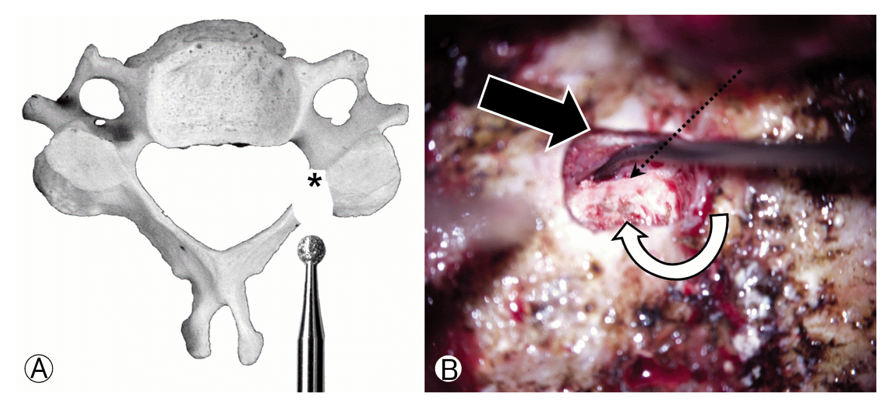
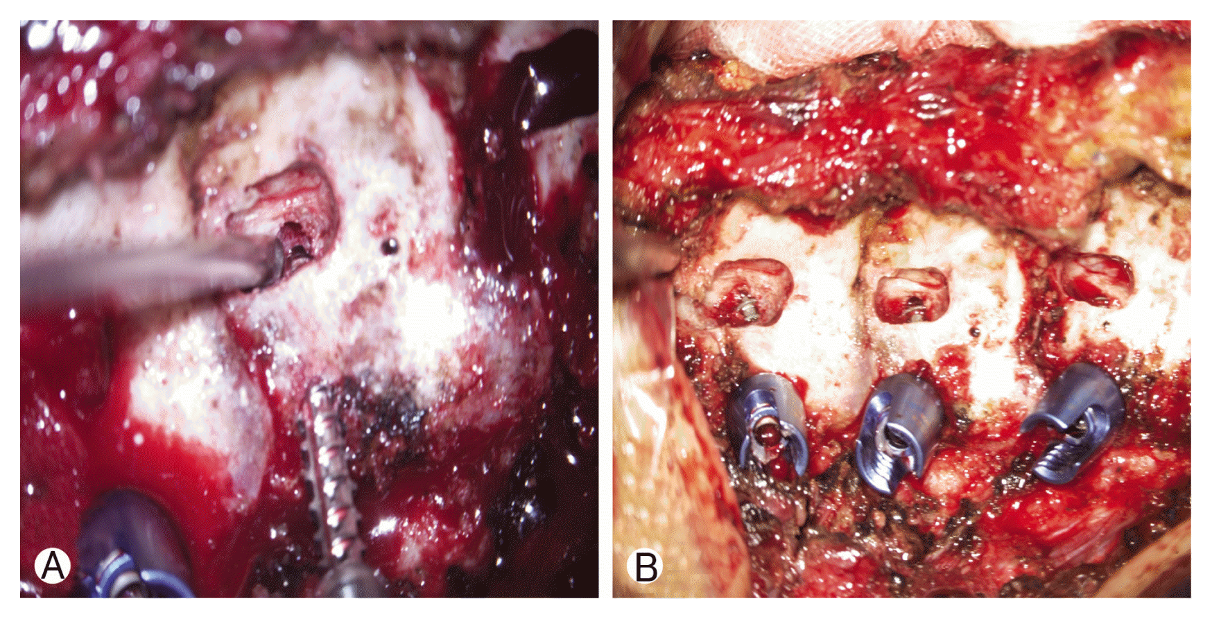
The lamina and medial portion of lateral mass were drilled with 2-mm burr under a microscope. After small laminoforaminotomy was made on the upper part of lamina, the ligament flavum at each level was gently dissected free from the inferior aspect of the superior lamina and from the superior aspect of the inferior lamina so that the arch of the medial pedicle cortex could be identified. After drilling the transition zone from lateral mass to the pedicle, the cancellous core of the pedicle was exposed. It was located at the medial part of lateral mass surrounded by the lateral portion of the pedicle (Fig. 1). Thereafter, the screw entry point was created on the superolateral quadrant of the lateral mass under lateral fluoroscopic guidance. Appropriate convergence angle was then confirmed via the exposed cancellous core (Fig. 2). After making an imaginary track from the entry point to the decorticated pedicle cancellous core, a screw was inserted via the decorticated site of medial pedicle cortex under direct visualization (Fig. 3). A trajectory was made so that the thread of the screw was exposed through the pedicle cancellous core. If the screw did not pass through the decorticated core of the pedicle, we changed the trajectory. If laminectomy or foraminotomy for neural decompression was necessary, these performed using high speed drill after pedicle screw insertion was completed. Bone fusion was performed using cancellous allograft.
3. Clinical and Radiographic Analysis
We reviewed the clinical records and radiographs of all patients. Clinical assessment included neurologic status and pedicle screw-associated complication such as vertebral artery injury and neural injury
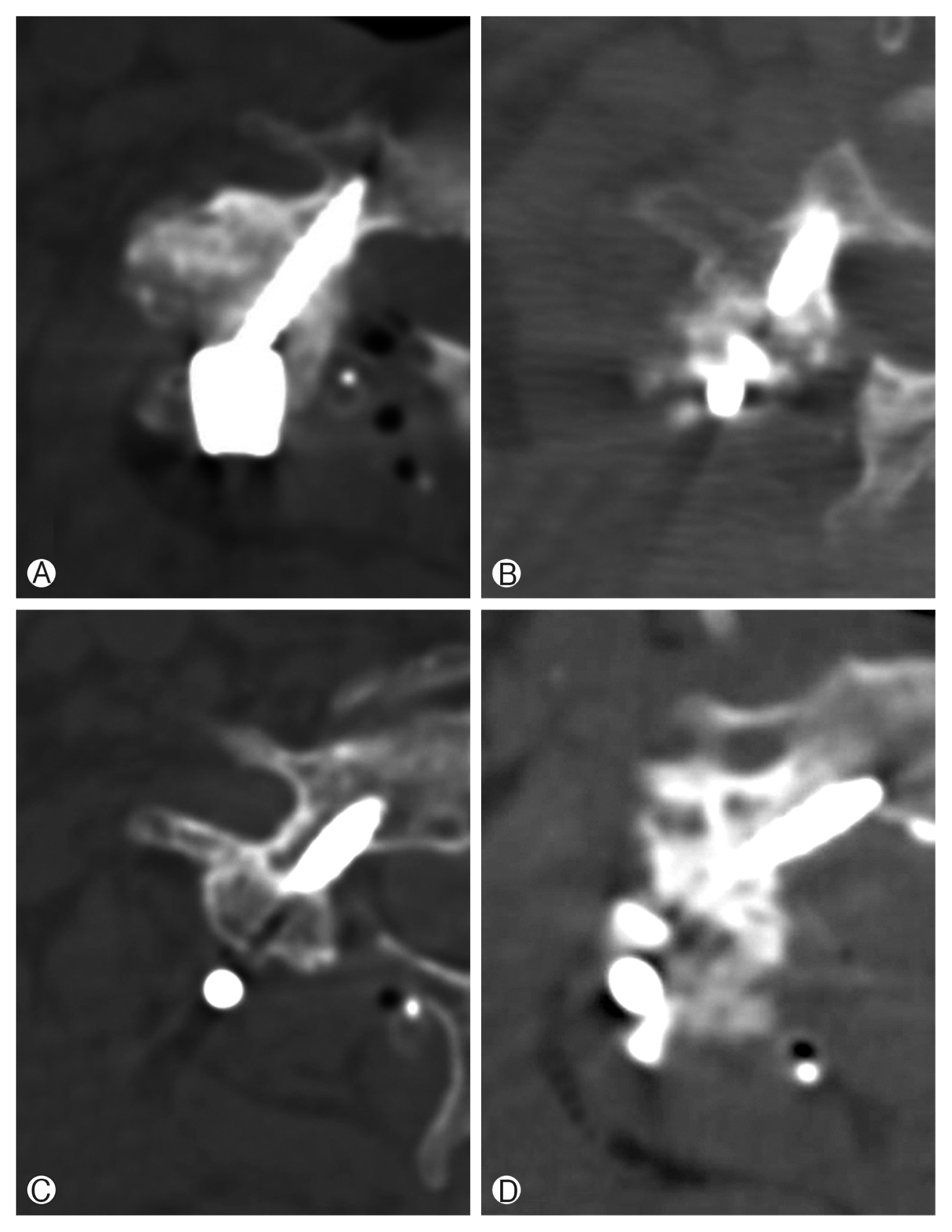
Axial computed tomography (CT) scans were obtained with 1-mm slices. Coronal and sagittal reformations were performed preoperatively and postoperatively to assess pedicle screw instrumentation. On preoperative CT scans, the convergence angle and the minimal diameter of the pedicle were measured. On postoperative CT scans, the degree of perforation was classified into four grades. If the CPS located within the pedicle and did not perforated, that was defined as grade 0 perforation. Grade 1 perforation was defined if perforation was less than 25% of the screw diameter. If perforation was over than 25% and less than 50% of the screw diameter, it was classified as grade 2. Perforation over than 50% of the screw diameter was defined as grade 3 (Fig. 4). Grades 0 and 1 were regarded as the correct screw position. Grades 2 and 3 were regarded as incorrect screw position. The direction of perforation was classified as medial, lateral, cranial, and caudal.
4. Interobserver and Intraobserver Error Analysis
The degree of perforation was confirmed 3 times by 2 junior neurosurgeons. If the degree of perforation differed between 2 junior neurosurgeons, the final decision was made by a senior neurosurgeon. Statistical analysis was performed using IBM SPSS ver. 18.0 (IBM Co., Armonk, NY, USA). The intraobserver and interobserver agreement rate and ╬║ values were determine the error between 2 observers who graded the pedicle perforation.
RESULTS
The interobserver agreement rate was 92.7% for the grade of pedicle perforation (mean ╬║=0.78). Intraobserver agreement rate was 91.3%(mean ╬║=0.71). The intraobserver and interobserver error analyses showed good agreement between the 2 observers. There was no conditions related to vertebral artery injury. Symptom related to nerve root irritation was not observed either after CPS instrumentation. None of the patients showed neurological deterioration after the surgery. There was no serious complication such as surgical site infection.
The average operative time was 174 minutes (range, 111ŌĆō247 minutes) and the mean blood loss was 234 mL (range, 100ŌĆō2,200 mL). A total of 88 CPSs were instrumented, of which 54 CPSs (61.4%) were classified as grade 0 and 29 (33%) were classified as grade 1. Four (4.5%) were classified as grade 2 and 1 (1.1%) was classified as grade 3. The mean pedicle diameter was 5.53┬▒1.15 mm, and minimal pedicle diameter was 2.76 mm. Mean pedicle angle was 41.4┬░┬▒11.1┬░. Ten patients needed laminectomy and foraminotomy for decompression. There was no facetectomy for deformity correction.
These CPSs were divided into correct position (CP) and incorrect position (IP). They were also divided into nonperforation group (NG) and perforation group (PG). Grades 0 and 1 CPSs were allocated into CP, while grades 2 and 3 CPSs were allocated into IP. NG was defined as grade 0 CPSs, while PG was defined as grades 1, 2 and 3. The overall rate of CP was 94.3%(83 of 88). IP occurred at C3 in 1 case, C4 in 2 cases, C5 in 1 case, and C6 in 1 case. IP did not occur at C7 (Table 1). A total of 34 CPSs were included in PG. In PG, 30 CPSs were classified as medial wall perforation while 4 CPSs were classified as lateral wall perforation. Superior or inferior perforation was not identified (Table 2).
DISCUSSION
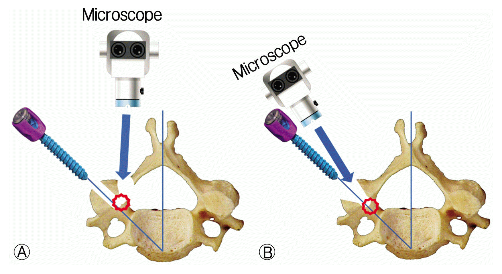
Medial funnel technique is basically similar to the funnel technique because they share the same landmarks: medial pedicle cortex with the cancellous core. Direct visualization of the cancellous core of pedicle gives confidence to the surgeon about the pedicle anatomy, and this leads high accuracy of CPS placement. Seo et al.19) have reported that the funnel technique is more accurate than the free-hand or the laminoforaminotomy technique in their cadaveric study. The difference lies in how to expose the cancellous core. In case of the funnel technique, the outer cortex of the lateral mass over the pedicle entrance is removed7). In the medial funnel technique, the outer cortex over the pedicle entrance is preserved while the cancellous core is exposed in the middle of screw path by laminoforaminotomy (Fig. 5). In addition, the screw entry point is made minimally on the superolateral quadrant of the lateral mass (Fig. 2). Therefore, the cortical bone near the screw entry point could be preserved maximally, thus maintaining the screw pull-out strength while maintaining high accuracy.
One of the benefit of this technique is that surgeons could see the orientation of pedicle structure without fluoroscopy or a navigation system. Surgeons could draw the imaginary axis of pedicle extended to the lateral mass surface through laminoforaminotomy, and expose the cancellous core of pedicle. This could decrease the necessity of fluoroscopy during screw instrumentation and reduce radiation exposure to surgeons.
The accuracy of CPS placement varied in literature, ranging from 16.8% to 97%4,7,14,19). Our series showed a relatively high rate of correct screw position (94.3%). This result was comparable to CPS placement with navigation system showing accuracy of 93.9% to 98.8%3,10,18,21). However, the navigation system has some limitation. At first, it is impossible for all institutes to have the navigation system because of the cost23). Second, there are some clinical conditions in which the navigation system cannot be used such as severe trauma cases requiring intraoperative reduction23). Finally, because the cervical spine is highly mobile, cervical spine alignment can easily change depending on the surgeonŌĆÖs force required for screw insertion22). It leads inaccurate synchronization to preoperative images. Therefore, some authors did not use navigation systems in CPS instrumentation22). Additionally, it takes long time to register images during surgery.
The pedicle architecture of the cervical spine usually leads to lateral perforation of CPS during pilot hole preparation, tapping or screw insertion15,17). Because the lateral pedicle cortex is commonly thinner than the medial cortex, the use of a blunt pedicle probe is usually directed toward lateral cortex7,15). This also increases the chance of lateral perforation of CPS. Recent multicenter studies have reported that misplaced screws over two-thirds are classified as lateral perforations when the conventional free-hand technique is used2,6). In laminoforaminotomy technique, the rate of lateral perforation has been reported to be from 72.4% to 74%4,14). However, the current study indicated that the medial funnel technique had a lower rate of lateral perforations (11.8%) than the conventional free-hand technique, the laminoforaminotomy technique or the navigation system for CPS placement2,4,19,21).
Our technique has some disadvantages. This technique needs partial laminectomy and process to find pedicle cancellous core. Therefore, additional operative time may be necessary. The risk of postoperative epidural hematoma or cerebrospinal fluid leakage may increase. In addition, intraoperative cord or root injury may happen. Uncontrolled epidural venous bleeding might also disturb the exposure of pedicle.
This study is also limited due to the relatively small number of screws and a heterogeneous group of patients. In addition, our results cannot be compared directly to other studies because the criteria for assessing development and degree of pedicle perforation were different according to each study. In this regard, uniform criterion for pedicle perforations and a larger multicenter study involving multiple surgeons with comparable patient groups are needed.


