



Abstract
The crowned dens syndrome (CDS), also known as periodontoid calcium pyrophosphate dehydrate crystal deposition disease, is typified clinically by severe cervical pain, neck stiffness and atlantoaxial synovial calcification which could be misdiagnosed as meningitis, epidural abscess, polymyalgia rheumatica, giant cell arthritis, rheumatoid arthritis, cervical spondylitis or metastatic spinal tumor. Crystalline deposition on cervical vertebrae is less well known disease entity and only a limited number of cases have been reported to date. Authors report a case of CDS and describe the clinical feature.
-
Keywords: Neck pain, Calcium pyrophosphate, Axis, Computed tomography
INTRODUCTION
Firstly, the crowned dens syndrome (CDS) was named by Bouvet et al. in 1985 as it was presented by cervical pain due to deposition of calcium pyrophosphate dehydrate (CPPD) crystals or hydroxylapatite at cervical spine and shown radiologically as crown or halo-like distribution when radio-opaque density appeared at the top and side of the odontoid process
4). CPPD crystal or basic calcium phosphate crystal is originated from hyaline cartilage which contains hypertropic chondrocyte, and it can arise from any joint that is composed of cartilage but rarely from tendon or bursa. If it occurs at cervicoaxial joint surrounded by several ligaments and if it causes severe pain and limitation of neck rotation at the same time, so it can be named as CDS
3,
13). Authors report a case of CDS and review of the literature of this underrecognized condition.
CASE REPORT
A 49-year-old female visited the outpatient clinic with a 7-day history of acute onset severe posterior neck pain. On the day of symptom onset, she took an acupuncture and chiropractic manipulation of cervical spine which did not turn out to be helpful, and she experienced gradual worsening of pain and marked restriction of neck motion. There was no history of trauma and medical illness. On physical examination, passive cervical spine movements were significantly reduced without any focal neurologic deficit. Range of neck motion was limited to 20 degrees on rotation whereas extension and flexion were slightly diminished. At the end of the range of neck motion, pain was markedly provoked. Patient's vital signs were stable.
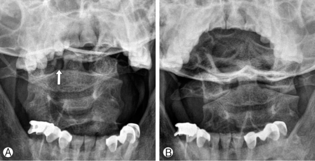
Plain radiographs of the cervical spine with open mouth view revealed abnormal radio-opaque lesion on the right side of the odontoid process (
Fig. 1A). There was not any evidence of spinal instability on dynamic images. Computed tomography (CT) images showed atlantoaxial synovial calcifications in a crown or halo-like distribution around the odontoid process (
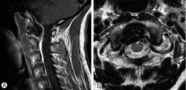
Fig. 2A). Magnetic resonance image (MRI) showed low signal intensity around the odontoid process lesion on T1 weighted image but not involving itself (
Fig. 3). Bone scan demonstrated increased radionuclide uptake around the odontoid process. We could not find any other lesion causative of pain within the spinal cord, nerve roots, intervertebral discs, cerebrospinal fluid and muscles. Laboratory tests revealed elevated high sensitive C-reactive protein (hsCRP) of 1.97mg/dL (normal range: 0.01-0.29mg/dL) while others were unremarkable. By integrating the result of tests listed above, we presumed a diagnosis as crowned dens syndrome.
Combination of prednisolone and nonsteroidal anti-inflammatory drug (NSAID) was taken and tapered off within three weeks, which led to resolve symptoms and normalize hsCRP. Neckbrace was put on to relieve motion-provoked pain during hospitalization. The lesion disappeared on plain radiographs followed in one month, which gave us a diagnostic clue to exclude such a condition from degenerative arthritis of the region (
Fig. 1B).
DISCUSSION
The crowned dens syndrome is a rare disease entity which radiologically shows calcification at cruciform ligament around odontoid process and clinically presents acute cervico-occipital pain, fever, and neck stiffness accompanied by biological inflammatory reaction as mentioned earlier. In our case, clinical manifestations include severe posterior neck pain of acute onset, significant restriction of neck rotation and elevation of serum CRP. So that clinicians could make a confusion with spondyloarthritis around atlantoaxial joint area.
The duration of pain varies from few days to several weeks, the location can be from suboccipital area to inferior posterior neck, and the characteristics of pain are also diverse from mild neck discomfort to sleep-breaking severe pain
5). Most of the crystalline deposits are calcium pyrophosphate dehydrate (CPPD) crystals or hydroxyapatite crystal. There is no specific symptom when the crystal's size is small, but as it grows, it causes chronic cervical pain or spinal cord compression. Calcification can occur at not only cruciform ligament but also transverse, alar and apical ligament. Especially when CPPD crystal deposition occurs at ligamentum flavum, compressive cervical myelopathy can be presented
8,
16). Dirheimer et al. performed plain radiographs to 27 patients who had articular chondrocalcinosis but neurologically asymptomatic, and demonstrated that syndesmo-odontoid joint calcification was found in 12 patients (44%)
6). Constantin et al. performed CT scan to 21 patients with articular chodrocalcinosis, and found that 14 patients had calcifications at transverse ligament and 5 patients among the 14 presented neck pain
5).
CDS can be misdiagnosed as meningitis, epidural abscess, rheumatoid arthritis, polymyalgia rheumatica (PMR), giant cell arthritis (GCA), cervical spondylitis or metastatic spinal tumor
1,
15,
18). Especially, A. Aouba et al. emphasized that association with cervical stiffness and shoulder girdle pain or jaw claudication can be mislead to PMR or GCA
1), and Laborie Y et al. reported that chondrocalcinosis such as CDS can be included in the category of rheumatic disease from a wide-spectrum perspective. However, it should be noted that the pathophysiologic process has not been completely proven
9), and therefore, caution should be used in relying on such statements or findings. There are several pathophysiologic theories about CPPD deposition at atlantoaxial joint. First, Constantin et al. set forth that chondroid cell in fibrous cartilage structures like meniscus of knee can be found in transverse ligament around odontoid process, and the presentation of these cell causes the disease
5). Another theory is that fibroblast in cervical spine ligament transforms into chondrocyte and the transformation may cause calcification
7).
For diagnosis, CT scan focused on C1-2 region is gold standard to detect calcification in transverse, apical and alar ligament. CT is even more helpful for small sized lesion when three-dimensional reconstruction is possible
1,
15,
17,
18). CT scan seems to be superior to other diagnostic modality for ruling out CDS-like odontoid process fracture or Os odontoideum, and so forth. However, when CT scan is performed long time after the onset of symptom, calcification around odontoid process cannot be detected as it has been absorbed. So, CT scan should be done as soon as the symptom occurs
10,
20). The presence of calcification can be assumed through plain radiograph by radio-opacity difference around odontoid process, but there is limitation as anatomical structures around odontoid process cannot be clearly distinguished. MRI is also helpful in identifying compressed spinal cord or soft tissues like nerve root, tendon, etc. What is interesting is that performing plain radiograph systemically over other joints like knee or wrist even though there is no specific symptom might be helpful because there were many cases which involve joints other than atlantoaxial joint in the CDS patients group
19). We couldn't find any abnormal joint except for periodontoid region. Elevation of ESR and CRP due to inflammatory reaction is common, and our case also showed slight elevation of CRP as 1.97mg/dL which could be explained by time interval between symptom onset and blood sampling
11).
Surgical procedure is performed rarely to patients who are diagnosed with CDS, but Baysal et al. demonstrates that 1 among 17 cases of CDS patients who progressively presented neurologic symptom and was treated by decompression surgery. And under microscopic examination, CPPD crystal in fibrous tissue was found and chronic inflammatory change was presented
2). In our case, when treated by combination therapy of prednisolone and NSAID, clinical symptoms showed dramatic improvement within 7 days. Although it was not clearly found that the period between the administration of NSAID and complete clinical recovery as many articles did not precisely describe, Ziza JM et al. and Resnick et al. reported that symptom had resolved on the 4
th day of administration and complete recovery had been made in 3 weeks
14,
20). Reginato et al. reported that after the dramatic initial improvement within a week, there is a slow but persistent improvement in 3 to 5 weeks as the second phase, and it is the same at other lesions with microcrystalline joint. They also reported that colchicine seems to be effective, though not much as the effect of NSAID. There were debates about use of steroid, and low dose administration of steroid could be helpful to patients who have no effect from NSAID
12).
CONCLUSION
Authors present a case of CDS as a rare cause of posterior neck pain with limitation of neck rotation. We suggest that clinicians should be aware of clinical feature of CDS for avoiding unnecessary lumbar puncture or biopsy, and cutting down hospitalization period.
Soonchunhyang University Research Fund
NOTES
-
This work was supported by the Soonchunhyang University Research Fund.
REFERENCES
- 1. Aouba A, Vuillemin-Bodaghi V, Mutschler C, De Bandt M. Crowned dens syndrome misdiagnosed as polymyalgia rheumatica, giant cell arteritis, meningitis or spondylitis: an analysis of eight cases. Rheumatology (Oxford) 2004;43:1508-1512. PMID: 15316123.
- 2. Baysal T, Baysal O, Kutlu R, Karaman I, Mizrak B. The crowned dens syndrome: a rare form of calcium pyrophosphate deposition disease. Eur Radiol 2000;10:1003-1005. PMID: 10879718.
- 3. Beutler A, Rothfuss S, Clayburne G, Sieck M, Schumacher HR Jr. Calcium pyrophosphate dihydrate crystal deposition in synovium. Relationship to collagen fibers and chondrometaplasia. Arthritis Rheum 1993;36:704-715. PMID: 8489549.
- 4. Bouvet JP, le Parc JM, Michalski B, Benlahrache C, Auquier L. Acute neck pain due to calcifications surrounding the odontoid process: the crowned dens syndrome. Arthritis Rheum 1985;28:1417-1420. PMID: 4084331.
- 5. Constantin A, Bouteiller G. Acute neck pain and fever as the first manifestation of chondrocalcinosis with calcification of the transverse ligament of the atlas. Five case-reports with a literature review. Rev Rhum Engl Ed 1998;65:583-585. PMID: 9809363.
- 6. Dirheimer Y, Bensimon C, Christmann D, Wackenheim C. Syndesmo-odontoid joint and calcium pyrophosphate dihydrate deposition disease (CPPD). Neuroradiology 1983;25:319-321. PMID: 6646410.
- 7. Doherty M, Dieppe P. Crystal deposition disease in the elderly. Clin Rheum Dis 1986;12:97-116. PMID: 3013494.
- 8. Fye KH, Weinstein PR, Donald F. Compressive cervical myelopathy due to calcium pyrophosphate dihydrate deposition disease: report of a case and review of the literature. Arch Intern Med 1999;159:189-193. PMID: 9927103.
- 9. Laborie Y, Berthelot JM. Rhizomelic pseudopolyarthritis: update. Rev Med Interne 2002;23:518-532. PMID: 12108176.
- 10. Malca SA, Roche PH, Pellet W, Combalbert A. Crowned dens syndrome: a manifestation of hydroxy-apatite rheumatism. Acta Neurochir (Wien) 1995;135:126-130. PMID: 8748801.
- 11. Ogata M, Ishikawa K, Ohira T. Cervical myelopathy in pseudogout. Case report. J Bone Joint Surg Am 1984;66:1301-1303. PMID: 6490709.
- 12. Reginato AJ, Reginato AM. Disease associated with depositon of calcium pyrophosphate or hydroxyapatie. In: Rubby S, Harris EDJ, Sledge CBet al., editors. Kelly's Textbook of Rheumatology. ed 6. Philadelphia: W.B.Saunders; 2001.Vol 1: p. 1377-1390.
- 13. Reginato AJ, Schumacher HR, Martinez VA. The articular cartilage in familial chondrocalcinosis. Light and electron microscopic study. Arthritis Rheum 1974;17:977-992. PMID: 4139958.
- 14. Resnick D, Pineda C. Vertebral involvement in calcium pyrophosphate dihydrate crystal deposition disease. Radiographic-pathological correlation. Radiology 1984;153:55-60. PMID: 6089266.
- 15. Salaffi F, Carotti M, Guglielmi G, Passarini G, Grassi W. The crowned dens syndrome as a cause of neck pain: clinical and computed tomography study in patients with calcium pyrophosphate dihydrate deposition disease. Clin Exp Rheumatol 2008;26:1040-1046. PMID: 19210868.
- 16. Sato R, Takahashi M, Yamashita Y, Izunaga H, Sakamoto Y, Yamamoto M, et al. Calcium crystal deposition in cervical ligamentum flavum: CT and MR findings. J Comput Assist Tomogr 1992;16:352-355. PMID: 1592914.
- 17. Scutellari PN, Galeotti R, Leprotti S, Ridolfi M, Franciosi R, Antinolfi G. The crowned dens syndrome. Evaluation with CT imaging. Radiol Med 2007;112:195-207. PMID: 17361376.
- 18. Wu DW, Reginato AJ, Torriani M, Robinson DR, Reginato AM. The crowned dens syndrome as a cause of neck pain: report of two new cases and review of the literature. Arthritis Rheum 2005;53:133-137. PMID: 15696551.
- 19. Zapletal J, Hekster RE, Straver JS, Wilmink JT, Hermans J. Association of transverse ligament calcification with anterior atlanto-odontoid osteoarthritis: CT findings. Neuroradiology 1995;37:667-669. PMID: 8748903.
- 20. Ziza JM, Bouvet JP, Auquier L. Acute suboccipital neck pain of calcifying origin. Rev Rhum Mal Osteoartic 1982;49:549-551. PMID: 7123130.
Fig. 1Open mouth view demonstrates radio-opaque lesion beside the odontoid process (arrow) (A). Open mouth view checked in one month reveals pre-existing radio-opaque lesion disappeared (B).
Fig. 2Coronal CT scan at C1/2 level demonstrates atlantoaxial synovial calcifications in a crown- or halo-like distribution around the odontoid process (A). Axial CT scan shows calcified lesion on the right side of periodontoid space and subchondral erosion (B).
Fig. 3Sagittal (A) and axial (B) T1 enhanced magnetic resonance image reveal non-enhancing, non-invasive mass like lesion around the odontoid process.
Citations
Citations to this article as recorded by

- Evaluación por imágenes de la patología calcificante y osificante de la columna cérvico - dorsal
Camila De la Barra, Consuelo Gatica, Fernanda Blaskovic, María Francesca Castoldi, Holvis Dellien
Revista Chilena de Neurocirugía.2026; 51(2): 84. CrossRef - Crowned dens syndrome: a rare yet intriguing condition of the spine
EA Acar, FC Tasgöz, S Uslu, O Soysal Gündüz
Scandinavian Journal of Rheumatology.2025; 54(4): 314. CrossRef - Non-Infectious Spinal Diseases Mimicking Infection: A Pictorial Essay
Jungmin Lim, Sung Hye Koh, Min-Jeong Kim, Su Been Lee
Journal of the Korean Society of Radiology.2025; 86(6): 970. CrossRef - Crowned Dens Syndrome als mogelijke bijwerking van zoledronaat
W. EL BAOUCHI, J. PEETERS, G. MOORKENS, D. JENNES, G. STASSIJNS, V. MERTENS
Tijdschrift voor Geneeskunde en Gezondheidszorg.2025;[Epub] CrossRef - Pseudogout of the lumbar spine
Sneha R. Gupta, Sai Phani Sree Cherukuri, Mohamad Chehab, Kalyan Paudel, Christopher Haas
Radiology Case Reports.2023; 18(4): 1490. CrossRef - Diagnosis and Treatment of Calcium Pyrophosphate Deposition (CPPD) Disease: A Review
Sharon Cowley, Geraldine McCarthy
Open Access Rheumatology: Research and Reviews.2023; Volume 15: 33. CrossRef - Case report: crowned dens syndrome in a patient with seronegative rheumatoid arthritis
Gianfranco Ferraccioli, Romeo Zanardi, Elisa Gremese
RMD Open.2023; 9(2): e003017. CrossRef - Crowned dens syndrome: A rare form of acute neck pain and headache that can be misdiagnosed or missed
Songsong Jiao, Fei Dong, Ruijia Liu, Jian Huang, Qingqi Meng
The American Journal of Emergency Medicine.2023; 70: 209.e1. CrossRef - Imaging of the Craniocervical Junction: A Pictorial Review
Kris Mertens, Filip M. Vanhoenacker
Seminars in Musculoskeletal Radiology.2023; 27(05): 499. CrossRef - Case Report: Posterolateral Epidural Supra-C2-Root Approach (PESCA) for Biopsy of a Retro-Odontoid Lesions in Same Sitting After Occipitocervical Fixation and Decompression in a Case of Crowned Dens Syndrome With Brainstem Compression and Displacement
Patrick Haas, Till-Karsten Hauser, Kosmas Kandilaris, Marco Skardelly, Marcos Tatagiba, Sasan Darius Adib
Frontiers in Surgery.2022;[Epub] CrossRef - A Quick and Comprehensive Guide to Differential Diagnosis of Neck and Back Pain: a Narrative Review
Elisha Krasin, Haggai Schermann, Nimrod Snir, Adrian Tudor, Eyal Behrbalk
SN Comprehensive Clinical Medicine.2022;[Epub] CrossRef - Crowned dens syndrome in a patient with severe secondary iron overload
Luani Barge, Gauri Gogna
Internal Medicine Journal.2021; 51(9): 1549. CrossRef - A Case of Acute Neck Pain: The Crowned Dens Syndrome
Pius E Ojemolon, Ehizogie Edigin, Narender Annapureddy, Augustine Manadan
Cureus.2020;[Epub] CrossRef - Cervical myelopathy due to subaxial calcium pyrophosphate dihydrate (CPPD) deposition with simultaneous asymptomatic crowned dens syndrome: two case reports
Dong-Gune Chang, Jong-Beom Park, Ho-Young Jung, Kyung Jin Seo
BMC Musculoskeletal Disorders.2020;[Epub] CrossRef - Thinking beyond pannus: a review of retro-odontoid pseudotumor due to rheumatoid and non-rheumatoid etiologies
Junzi Shi, Joerg Ermann, Barbara N. Weissman, Stacy E. Smith, Jacob C. Mandell
Skeletal Radiology.2019; 48(10): 1511. CrossRef - The Crystal Crown
Hasan Ahmad Hasan Albitar, Delamo I. Bekele
Mayo Clinic Proceedings.2019; 94(8): 1641. CrossRef - Crowned dens syndrome presenting as pyrexia of unknown origin (PUO)
Agam Bansal, Mohit Gupta
Romanian Journal of Internal Medicine.2019; 57(3): 266. CrossRef - What a pain in the neck: Crowned dens syndrome as an uncommon cause of neck pain
Alexander M Pandelidis, Alan W Dow
Case Reports International.2019; 8(2): 1. CrossRef - Elongated odontoid process in late Holocene skeletal remains from B6 archaeological site, Mendoza, Argentina
J.A. Suby, P. Novellino, G. Da Peña, C.D. Pandiani
International Journal of Paleopathology.2018; 22: 86. CrossRef - Crowned Dens Syndrome: Calcium Pyrophosphate Deposition Disease Masquerading as Osteomyelitis
ALEXANDER HECK, NATHANIAL NOLAN, CHRISTIAN ROJAS-MORENO
The Journal of Rheumatology.2018; 45(10): 1422. CrossRef - Dr. Stein’s Problem-Based Learning “I Can’t Walk Because of Fever and Physical Pain!”
TOSHIO NAITO, YUKIKO FUKUI-WATANABE, YUKI UEHARA, MAKOTO AOKI, GERALD H STEIN
Juntendo Medical Journal.2018; 64(5): 338. CrossRef - Acute neck pain due to crowned dens syndrome - A case report -
Yeojung Kim, Youngkwon Ko, Wonhyung Lee, Yongsup Shin, Chan Noh, Seounghun Lee, Hyunwoo Park
Anesthesia and Pain Medicine.2018; 13(4): 435. CrossRef - Crowned Dens Syndrome
Kiyoshi Shikino, Takahiro Ota, Masatomi Ikusaka
The American Journal of Medicine.2017; 130(3): e111. CrossRef - Crowned Dens Syndrome Occurred in a Patient after Simple Drainage for Chronic Subdural Hematoma: A Case Report
Ryota Tamura, Satoshi Takahashi, Dai Kamamoto, Tomo Horikoshi, Kazunari Yoshida
NMC Case Report Journal.2017; 4(1): 15. CrossRef - Usefulness of cervical computed tomography and magnetic resonance imaging for rapid diagnosis of crowned dens syndrome
Akihiro Inoue, Kanehisa Kohno, Satoko Ninomiya, Hitomi Tomita, Shinji Iwata, Shiro Ohue, Kenji Kamogawa, Kensho Okamoto, Shinya Fukumoto, Haruhisa Ichikawa, Shinji Onoue, Saya Ozaki, Bungo Okuda
International Journal of Surgery Case Reports.2017; 30(C): 50. CrossRef - Elderly Male With Neck Stiffness and Fever
Taichi Imamura, Seth Lotterman, Catherine Glazer
Annals of Emergency Medicine.2017; 69(5): 665. CrossRef - Crowned Dens Syndrome Associated with Bowel Cleaning for Colonoscopy
Naoto Kohno, Yoichiro Kobori, Shuhei Yamaguchi
Internal Medicine.2017; 56(19): 2645. CrossRef - Crowned dens syndrome: a rare cause of neck pain and fever
Matthew Jiang, Sukumar Navanathan
Medical Journal of Australia.2017; 206(5): 199. CrossRef - Magnetic Resonance Imaging and Computed Tomography in the Evaluation of Crowned Dens Syndrome Secondary to Calcium Pyrophosphate Dihydrate
Viju Moses, Hemant A. Parmar, Amr H. Sawalha
JCR: Journal of Clinical Rheumatology.2015; 21(7): 368. CrossRef - Crowned Dens Syndrome as an Initial Manifestation of Crystalline Deposition Disease
Ryo Koda, Yohei Tsuchida, Kazutaka Yoshizawa, Kazuo Suzuki, Akio Kasai, Tetsuro Takeda, Junichiro James Kazama, Ichiei Narita, Kazukiyo Yoshida
Internal Medicine.2015; 54(18): 2405. CrossRef - Crowned dens syndrome
Ryota Inokuchi, Kazuma Ohshima, Miyuki Yamamoto, Tatsuma Fukuda, Kensuke Nakamura
The Spine Journal.2015; 15(6): 1499. CrossRef - Crowned dens syndrome
Taiki Yamada, Takeji Saitoh, Hironao Hozumi, Yoshiaki Takahashi, Masashi Nozawa, Toshiaki Mochizuki, Atsuto Yoshino
Acute Medicine & Surgery.2015; 2(4): 273. CrossRef